
Smarter Dentistry
AI and your practice
Transcript
Hello, everyone, and welcome to this webinar. Thank you so much for joining us today. This webinar is about SmarterDentistry, AI, and your practice.
I'm joined by doctor Nishan Dixit, who will introduce himself shortly. My name is Rabeha.
I am a customer success manager here at Pearl and also a dental hygienist.
So today's webinar is going to cover some of the things related to AI, of course. We're going to talk about the role of AI in modern dentistry, how AI applies to a clinical setting, and how AI can contribute to supporting your diagnosis and increase your case acceptance. So if I just pass it over to Nishan, would you kindly introduce yourself, please?
Hello. Hi. So as Rupia said, thank you very much for, allowing me to do this webinar.
And so I'm Nishan Dixit, and I'm a general dentist who has a special interest in minimal invasive aesthetic dentistry.
I qualified from Guy's Hospital in nineteen ninety four.
And thereafter, I did my BT straightaway in Northwest London and worked as an associate in another practice in Northwest London as well pretty much for five years, and then established and or bought, Blue Court Dental in two thousand, practice located in Harrow.
Initially, it was a single handed practice, and we, bought some property behind the practice and expanded it into, what is now a three surgery practice, with two hygienists working there as well alongside myself and my wife and the, the rest of the team.
So initially, when I was in practice, I was involved with an organization called the AOG, which is the Asian, Odontological Group Anglo Asian Odontological Group. Get that right. Sorry.
And I was involved with them for the best part of ten years in various capacities.
Then once I'd finished my term and my roles with those, I moved on to the BACD and initially joined, the committee's, education committee, and then moved up, to the board of directors and was the scientific director for, very few years, five, six years or so, and eventually became the leadership team and and moved on to becoming president in twenty nineteen, twenty twenty.
After BACD, I'm currently only involved in, one other thing now, and so I'm a member of the GDP advisory board for AI, and I have been for the last five years or so.
And recently, over the last couple of years, I was involved with, another organization called Safe Smiles where myself and a few other key members for leading organizations, dental organizations, were trying to involve were involved in trying to help patients choose correctly rather than having teeth, smiles treated AI, mail order companies.
So, basically, what is artificial intelligence?
AI has been around for many, many years. Pretty much started in the fifties by, Alan Turing.
And, he was involved in the, he used a machine, test called the, imitation game. Sorry. Imitation game. And he this was the first machine intelligence test that was carried out.
So AI is simply a set of algorithms that work together, to accomplish goals set by humans.
Computer AI are involved in training these algorithms.
They feed them troves of data, by a process called machine learning.
These algorithms, they learn to recognize patterns of in information, which are ultimately allows the AI to accurately, categorize these and make predictions about how we show it.
So what is artificial intelligence?
AI has been around for many years, pretty much since the fifties, and was started by, Alan Turing.
And he was, instrumental in in developing this, and they had a system called the imitation game, which is basically machine intelligence.
It's a test machine intelligence test that's carried out, and that's what led to the the birth of AI.
It's been used in medicine and dentistry for ages as well. Oh, sorry. Medicine for a for a while now. But AI is is, to put it simply, is a set of algorithms that work together to, to work together to accomplish goals set by humans.
Computer scientists train these algorithms by feeding them troves of data. This this sort of process is called machine learning.
And the algorithms learn to recognize the pattern in information which ultimately allows, the AI to accurately categorize and make predictions about how we show it.
So like I said earlier, it's been around in in medicine for a bit of time, and it's been used in the heart, the brain, and the lung scans.
So why can't we use it in dentistry? You know, it's used accurately and for for precision and, and speed in the in medicine. So let's bring it to dentistry.
And and so Pearl did. And Pearl have is one of these companies that use this technology successfully and are, you know, rolling it out to dentists.
But how did I get involved?
So for me, I got interested a few years back when I went to the Chicago Midwinter Conference, and I saw Pearl, exhibiting there along with other companies similar companies.
I had a few demos and was really kind of wowed by it, and and I thought this would be perfect for my practice and my patients.
Eventually, you know, been, that I believe that we're a progressive practice. And I'd like to think we wanna keep improving our patient experience, our patient service, patient journey.
So I thought we we needed to to get involved with Pearl, and bring Pearl to our practice.
And and setting ourselves apart from other practices is is the key to moving forward, I believe.
So just a little bit about the the x-ray evolution.
So when I first started dentistry in ninety four, we had the the old film x rays and, you know, it was interesting, you know, at the time, you would use a, automatic processor, with, fix, fixers and developers in in the machine, and you'd come out with an x-ray pretty much as you can see on the left hand side.
Interestingly, before this, the forefront of this was the old darkroom. And when I actually bought my practice in two thousand, the previous practitioners actually had a dark room where he was processing the x rays manually and and hanging them on a, on some paper, so wire mesh type thing, and let them develop over a few days. And, when I saw the patients, for the first time, they were like, oh, we gotta come back to see our x rays. And I said, no. We've got an automatic processor. So they were a little bit, wow. You things can be seen in five minutes.
Next, the evolution went further and digital x rays came about.
And we actually bought a digital scanner in, We went digital twenty twelve stroke twenty thirteen and, changed obviously the the quality of the X rays, the consistency of the X AI.
And, again, going from five minutes processing now to seconds.
The the next logical step then was, AI technology to read those digital X rays. And and we we purchased Pearl, around about June, July and have been using it since to to great effect.
So from what we've seen is if you look at the the manual x rays, you know, we were throwing them up and the patients look at these x rays. We're we're fortunate.
As clinicians, we've had the, the knowledge. We've been we've studied at uni and had further training on x rays and how to interpret them. Our patients just see black and white.
They don't know. They they kind of go without judgment and you build up that trust over a period of time.
But but the reality is that according to the data, sixty five percent patients really don't understand what they're looking at. They're nodding their heads when you're telling them something's wrong.
Of the of another fifty nine percent really don't trust what they're we've been told. And, you know, when you tell them you've got an air carious lesion, they'll be like, but nothing hurts doc. Or, you know, why do I need that treated? Why have I got a hole? I'm not having any pain, any signs or symptoms.
You just wanna drill for the fun of it.
And, seventy eight percent say trust in diagnosis is key for them to accept in treatment. So, you know, you build that trust up with the patient over a period of AI, but use adjuncts. And, you know, in the past, we've been using you got X rays, and then you couple that with interval cameras.
And then now we're using, interval scanners with, NHERI technology. So infrared technology, near infrared technology.
So that helps, show patients what's going on.
With the AI, added to this, it's just gonna, increase that level of trust. And you're using every single tool available to you to explain to patients what is going on in their mouths.
So here are a few points that I believe will transform, the way AI is used in your practice.
Some have become evident with what I've been doing over the last few months, and, you can just see things like improved accuracy, diagnostic accuracy.
I'll go through a lot more of these points as we go along, but, these are really more personal to what we've been doing.
And so if we start with the increased diagnostic accuracy, PERL can can detect many things from early caries lesions, established caries lesions, obviously, and bone loss. We've got calculus. We've got, deficient, restorations, pathology, ledges, calculus, so, eight core lesions as well.
So earlier detection and more AI more accurate diagnosis is is given to the patients by using this sort of technology.
You can use by by seeing things early, you can treat things early, and prevent the disease progressing further.
And, ultimately, that will always give a better patient outcome.
In as you can see from this X-ray that we took over a patient recently, there's a few things that we we see from the X rays and we can sit and talk to the patient about.
One is their bone levels.
And these these numbers represent, the amount of bone loss from the CEJ to the bone crest. And, where it's increased a little bit, we can explain to the patients why it's increased, where they need to focus on their cleaning.
And then on on top of that, you can see the pink lesion, and that's basically early caries.
So it detects helps detect early caries. You may not see it clinically, and it may not even be visible to the naked eye on on on an x-ray.
So once you've seen it, you can explain to the patient what's going on and and look.
Here is an area of early caries.
And our feedback has been that the most common question they ask straight away is, so what can I do about this?
You know, how can I how can we treat this? And, obviously, being a hundred percent into the enamel, we won't intervene, but we explain to them how they can, keep it clean, how they can do their best to to slow the progress progress of that caries lesion or even arrest it, with different methods.
Pearl has established or Pearl has, has really done a few studies and it's it's said about thirty seven percent more disease is detected, which is kind of synonymous to what we see in in our patients at the moment that we we are definitely seeing, seeing more, especially the early caries lesions. And, more importantly, on top of that, we are, getting more more patients coming back for other treatments as well, on what they've seen on the x rays. They they they trust the AI, so they're they're having their treatments done.
We all miss things in a hurry.
So, basically, with Pearl, things are AI, and, it will point Pearl will point you to areas that you're you've missed that may need further, looking at.
So with Pearl, again, a case scenario I've put up there, but we have a lot of consistency and there's a reduced human error in what we see and find on our X rays.
Taking a couple of AI wings, you areas are AI, the caries lesions and, late, caries lesions. So so this this X-ray that I put up is obviously, the same bitewing, right handed right sided bitewing showing two things. One is the early decay, which is a hundred percent into enamel, but another one showing, a late or progressed caries, which shows us about forty five percent into into the dentine already.
And then, you know, coupled with that, I explaining to the patient the difference between the two and, they are more you're more likely to take up treatment.
And the early lesions, you can monitor with X rays on a on a more regular basis, and see how that progress is has been made rather than guesstimate, you know, or eyeballing it as we used to do.
The the the pearl also reduces the chances of any missed areas, or any false positives.
We said interpretations, you know, so the results are more reliable and more accurate.
Enhanced workflow efficiency.
So for us at our practice, we found that, the the the whole patient journey has changed with with Pearl. We we find that, I can by having Pearl in the practice, it speeds up my diagnosis process so I have more time focusing on the patient care rather than spending time reading looking at X rays. Obviously, I will look at X rays and scan them first and then, use the Pearl software, and and it will highlight areas where I need to go to straight away. And and that allows me to spend more time talking about these lesions with the patient rather than wait then waiting in the chair while we scan through things.
So patient fair care is is the focus of our attention.
And the the the, ahead of the process of viewing an X-ray is streamlined.
So by streamlining that process, again, productivity is increases.
You have more time on your hands to to spend with the patient.
So with the cost effectiveness of, Pearl, I mean, you find that patients don't always understand what's going on in their mouth. You try your best to explain things.
But by visually showing them, what's going on, there is less chance of, misdiagnosis and delayed treatments.
So what we find is that a small lesion, you explain to a patient they're more likely to take the treatment on, and hence will be less costly for that patient. Or if you if you're not seeing it on your own x rays, you'll AI, you know, that lesion progresses into something more, a larger restoration problem. So it saves the patient time and money, and also, gives you more time to focus on a on other aspects.
The patient isn't coming back for more treatment or more lengthy treatment.
So with we we find with our patients that, you know, all the patients now are are very much tech savvy. They are they see things on social media. They read Google.
So they know what's out there. They know what's available.
They do expect your, practice to be high-tech now, and they want, state of the art health care solutions and treatments.
They want a practice that has up to date digital x rays. They want patients that oh, sorry. They want, practices that have scanners, that can look at, you know, your teeth and look at the wear on your teeth, look at lots of lots of things in for cameras to see what's going on.
So Pearl is another tool, and you'll find that once the patients have this, visual aid, they they start, their trust and satisfaction with yourself is is increased.
It shows that you're you're committing them. You're committing your practice is committed to, you know, top tier, level of care.
They they appreciate you know, we found our patients have have really appreciated the speed and the precision of the software, and they they really do trust and their satisfaction has improved. You know, they have our feedback from over the last few months has been that our patients are are highly impressed that we are keeping up to date with new technology and and investing in their their own health.
That's really the the best feedback we've had so far that the the are wowed by what we have on offer and what Pearl can do for them as well.
So this this data really we saw the pearl have, have found is is from a thousand US consumers.
So it is a bit American centric, but it does apply to the UK as well.
Sixty three percent of patients are really uncertain if their dentist is reading their x rays accurately, you know, and that goes down to them not understanding what they're seeing.
Fifty five percent of them would feel more confident if another dentist verified their x rays, especially when you say to someone you've got, you know, decay in your mouth and they've been told, there's nothing wrong.
So there is that trust has to be gained in in other ways and visual aids is the main thing, I think, so they can understand.
Though we've got to remember that patients are not trained to read x rays.
They you have they have to trust you.
And this is using pearl as an an added way to gain their trust.
Eighty two percent of patients, would prefer that their dentist use, more advanced AI technology and die and diagnostic aids in, helping them understand their x rays.
So with scalability, this this is kind of, for those principles that want to buy more practices, so ideal tool to have, because two things really, I feel. But one is that you've got associates who can help and diagnose and treat and plan, cases. You've got a a lot more consistency between clinicians. So what a practice with multiple clinicians on-site, the the accuracy, is the same. We all see x rays and we all read things differently on an x-ray, but Pearl doesn't. Pearl is the same for every case, for every clinician using Pearl. So there is consistency with, what what is being found and what is being interpreted from the x rays.
The patients are not being told different things.
They have, consistency with, their report the reporting on their x rays.
Obviously, the software itself can handle, increased amounts of data and patient volume without without requiring, additional staff.
My my belief that is that AI is the future of dental care.
We've been using AI for multiple things. AI is used in, our note keeping.
We use it within our scanning.
There are even, telephone systems out there that will, record the conversation and put down a summary of it in a word document instantaneously.
So AI is definitely the future of dental care. So why not bring that, region of our x rays as well?
Adopting the software now helps you prepare your practice for the future and future innovations, allowing you to stay competitive in the market.
As the technology advances, I feel early adopters will be in a better position to integrate new features and technologies further will improve the practice performances.
So looking at some data, again, it's a little bit US, based, but some of these figures are, are quite interesting really. So looking at the first one, forty six percent increase in treatment volume for restorative fillings, restorative work, and twelve percent increase in in hygiene.
That's, for me, that's bread and butter dentistry.
So Pearl is helping you increase your production by highlighting areas that you you may have missed from your X-ray AI, but also it helps, with the patients understanding what's going on in their mouth and more treatment is probably taken up. We have definitely found an increase in our treatment uptake since using Pearl.
Patients understand more about their their mouths. They understand what they're seeing on the x rays, and they literally their question is when can I get booked in to get this done?
And that's the bread and butter. Coupled that with further treatments that may be needed down the line, crowns, implants. They can see the angulation. They can see space closures.
There's a lot more they can understand from a color X-ray than from a black and white X-ray. And Pearl for me is is perfect for this.
Back to confidence and trust.
We we've we've seen a we've been seeing a lot of more patients since COVID.
A lot of patients are coming back to us after four or five years who haven't been worried about COVID. COVID was always on their mind.
Now when they've come into our practice, their focus is more about AI.
Older practices are now using new technologies, got AI, and, they really, really are impressed with that. They they see things, as I've said, they're being they they are more tech savvy. They they expect they've heard about AI in in various guises, and then you tell them we're using it as well in X rays, and it's like, wow. Well, this is amazing.
You know, we find that, seventy one percent of patients you well, not we AI.
Pearl have found that seventy one percent of, patients will trust the dentist diagnosis if backed up by AI. I mean, that that speaks volumes because there's obviously a little bit of, patients AI unsure about what is needed. They are they they come into the dentist knowing that there's not they feel that nothing's wrong in their mouth, and then they've been told they need one or two restorations, etcetera.
This this just backs up what they're seeing. Visually, they can see what's going on, and you can you can demonstrate that using the software.
I think using the software, the patients are more assured. Definitely, our patients feel, they are more assured, that using by using software, there is more accuracy, in diagnosing what's going on in their mouth, and, their quality of treatment seems to be, what is necessary what is important to them. They want they want the best, and we're giving them the best as we can with all the technology that's available.
So with compliance and documentation, Pearl Pearl has, we all have to when we take, sorry. Let me backtrack. Pearl, we when we take X rays, we have to document our findings. We have to report on the X rays.
And Pearl kind of helps with this process. It complies with the the legal requirements and the industry standards. There is a button on on the Perl software app that will allow you to to do a printout of what you AI.
So it will AI.
Sorry. Pearl Pearl will actually show you the the lesions that are there, the levels of bone, etcetera. And you can print that out, attach it to your patient records, attach it to your treatment plan, and you're complying with, the requirements.
Obviously, the, records, sorry, the the records that are kept, you can, compare the records over a period of time, AI, x rays if you've got lesions that are small a year later or eighteen months later when you take the next set of x rays.
Those those measurements can be compared and you can see if there's a deterioration in those and does it need intervention, does it not need intervention.
One other way or the best way for me to really go through compliance legal requirements, I think, is to go through a case study.
A real life patient that came to see us in August.
He was he'd seen been seen at another practice a couple of months prior to this, had an examination, a couple of AI wings, and, a quick clean, was told nothing was wrong.
A bit bit of his history was that, he had a restoration done on the lower left six six six months ago and it kept leaking. And I think he had it repaired two, two or three times since.
He was recommended to see me for some possibly some other work that he was interested in. But at the time he came in to see me, the the lower six was giving him a few problems, and he was told nothing was wrong by his previous dentist.
The problems were the occasional sensitivity with hot and cold. Previous dentist had just said, look. Everything's fine. X rays are fine. Just use some.
It all settled out.
Looking at the X rays, we we obviously requested these X rays from the previous practitioner.
And and looking at them, you could probably see a couple of lesions on the the premolars if you look closely. I know the accuracy or the quality of these digital X rays are not great, but even on on this poor quality, you can make things out and the, deficiency of the lower six restoration.
We explained, we explained the, the deficiency on the six. You could see it was clinically it was also breaking down, and it needed some intervention.
We went through, his case with him and said to him that, look. Once, we'll restore the six.
We, once we got the X rays, I called him back for a second, consultation, and we put the X rays into Pearl.
And a few things showed up. We we highlighted the the, calculus that was visible was was there, And then we went through all the pink dots with him. The early caries lesions first, explained to him that these are early. They don't necessarily need intervention.
But some of the other the other two lesions did need intervene intervening, and he was booked in, for the the restorative work. We put it into the the color as well, and you can quite clearly see that some of these lesions had gone into dentine and needed active intervention. And the the six, he could quite clearly see the the deficiency in the restoration.
So the treatment took, two or three visits. We restored him. He had some hygiene work done, and and now he's currently in the middle of some Invisalign.
Very happy. And the six is stabilized.
It just needed a simple intervention.
A composite respiration was done for him.
If we'd have left that, you probably would have found that may have progressed or deteriorated into something larger. So early intervention, or a timely intervention was the key.
Some of the other x rays we took of this patient as well, you can see the OPG, again, PEARL. You can import the OPG into PEARL, and it will show the caries lesions. It will go through some of the anatomy quite nicely, and and it's all things you can explain to the patient. And it looks all very pretty as well. The patients can understand things very clearly.
Pretty much, a a sense of AI diagnosis is really important.
So in in summary sorry. In summary, we have a a few things. Pearl Pearl's great at diagnosing or or lead guiding you of where lesions are. It will tell you there are early caries lesions. It will tell you that there are late caries lesions.
Periapical radial lucency is highlighted.
Calculus, which means a hygienist can focus on areas where she he or she is needed to clean and the patient is needed to clean those areas.
Possibly, periodontal treatment further periodontal treatment may be needed.
Bone levels are monitored. You can overlap these bone levels with x rays when you take them in the future and see if there is any deterioration.
And the patients can understand that more rather than visually just showing that there might be a little bit more bone loss than normal. You you've got a bit more of an accurate accurate measurement to say that, you know, you you're beginning to lose a bit more bone here. We need to intervene more or we need to refer you on further, so on and so forth.
Restorations can be monitored.
Ledges or margins, you know, we can intervene a lot earlier rather than let that things deteriorate and suddenly a root canal develops.
So when we when we qualified, we were all told one thing.
You can't treat what you can't see.
So get some loops and maybe, an additional light as well.
Now I think the focus is slightly slightly different as well. You can't treat what you can't AI.
So maybe we need to get Pearl as well to help us with this.
Thank you for listening.
Any questions?
Thank you, Nishan. That was very interesting.
Thank you to the audience. You will have some CPD questions to answer to be able to obtain your CPD certificate.
We'll leave this up for a little while so you can grab a screenshot or access the website to get a demo booked.
Have a lovely evening. Thanks, Nishan.
Thank you AI.
Meet Your Host
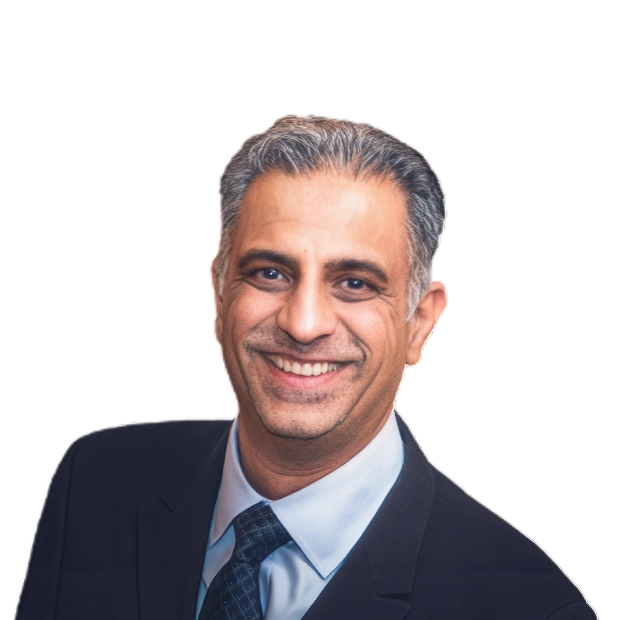
Dr. Nishan Dixit graduated from Guys Dental Hospital in 1994 and spent the first five years of his career as an associate in various practices across North-West London. In 2000, he founded Blue Court Dental in Harrow, Middlesex, where he provides comprehensive dental care while specializing in minimally invasive aesthetic dentistry. Dr. Dixit has been a member of the Invisalign GDP Advisory Board for over five years and is a past president of the British Academy of Cosmetic Dentistry (BACD), where he served on the board for 11 years, primarily as the scientific director. Additionally, he spent a decade on the Anglo-Asian Odontological Group (AOG) Committee in various roles. His commitment to advancing his skills has led him to train with leading clinicians both nationally and internationally.
Done learning and want to get started?
Book a demo