Simpler, Faster CBCT Review for Everyday Care
FDA-cleared AI for CBCT that transforms complex scans into clear, actionable 3D insights so every clinician can review, refer, and plan with confidence.
Get started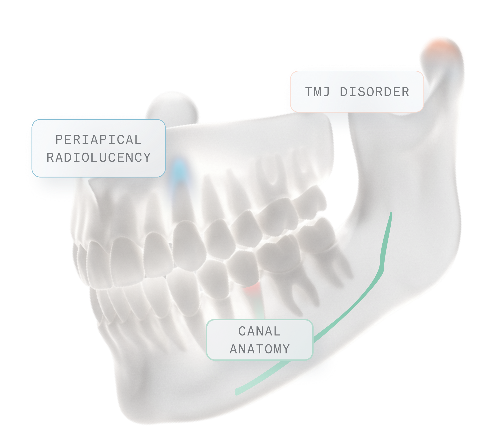














First-Ever FDA-cleared AI for 2D & 3D Dental Radiology
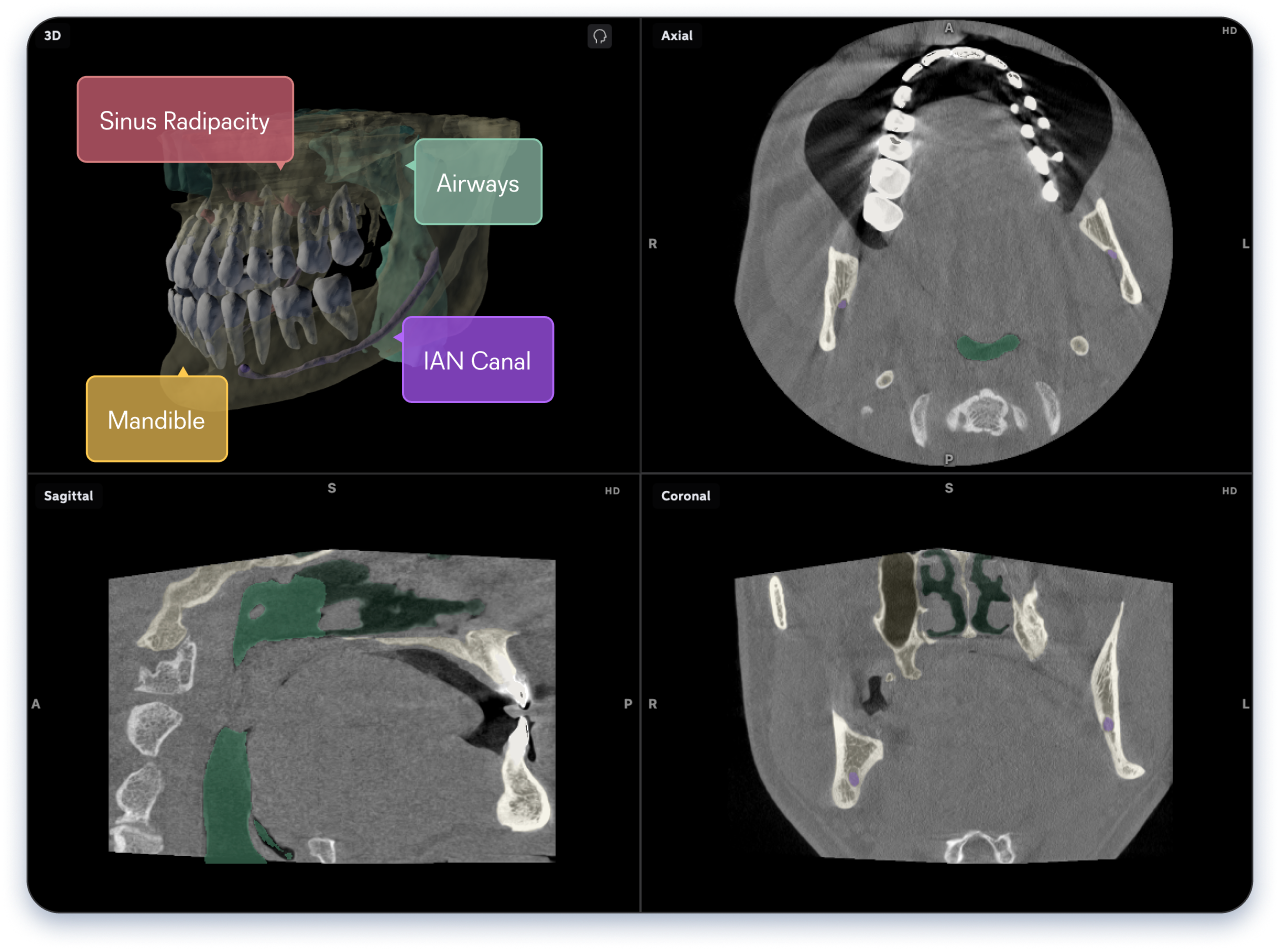
Transforming hundreds of slices into intuitive, color-coded 3D structures, Second Opinion 3D gives clinicians deeper insight into 3D data in minutes — not hours.
3D Clarity for Every Clinician
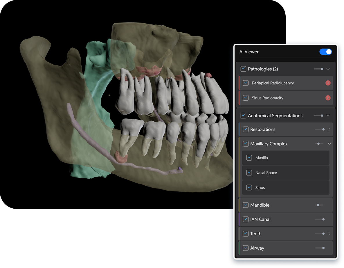
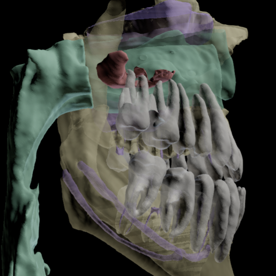
Pearl color-codes and segments key oral and maxillofacial anatomy, while AI findings surface clinically significant pathology* that demands attention.
- Clean, annotated visualizations make complex 3D data digestible and actionable
- FDA-cleared anatomical segmentation color codes key oral-maxillofacial anatomy
Expert Radiology and Reports, No Extra Work
Send images to licensed radiologists and get insurance-compliant imagery with just one click.
- Request referrals for identified asymmetries, lesions, and anomalies
- Support requests with labeled, color-coded 3D visuals and clinical notes
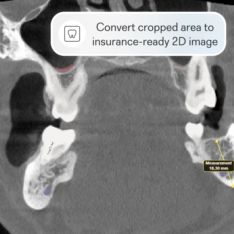
- Export CBCT imagery in 2D formats that insurers can review
Automatic Sync
One-Click Referrals
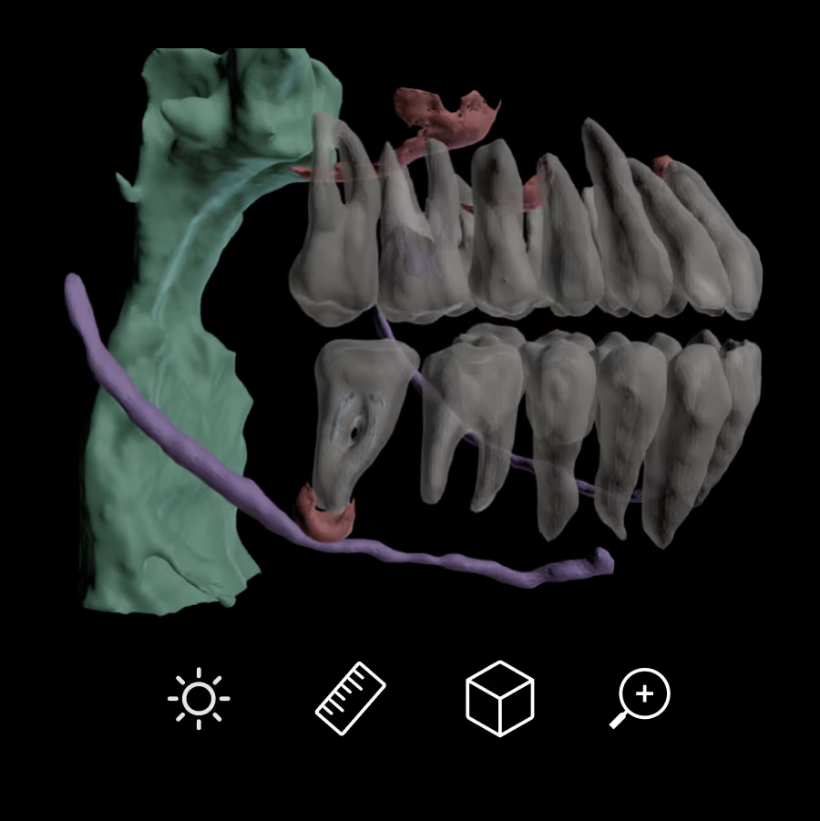
Interactive 3D Viewer
Fast, Intuitive Workflow

Book a Demo
Pearl’s newest FDA-cleared innovation brings real-time AI to CBCT imaging, adding a new dimension of radiologic clarity to dental imaging.
FAQ
What is Pearl Second Opinion 3D® and how does it help dentists read CBCT scans?
Second Opinion 3D® is Pearl’s FDA-cleared AI for cone beam computed tomography (CBCT) analysis — making Pearl the first and only dental AI company with FDA clearance for both 2D and 3D imaging. It automatically segments key anatomy, including the inferior alveolar canal, maxillary sinus, nasal space, airway, mandible, and individual teeth, so dentists can review complex CBCT scans faster, interpret findings with more confidence, and explain them clearly to patients chairside.
Can Second Opinion 3D detect pathology on CBCT scans?
Yes. Second Opinion 3D includes AI-powered pathology detection covering temporomandibular joint disorder (TMD), carotid artery calcifications, impactions, periapical radiolucency, and sinus radiopacities. This helps providers surface clinically significant findings more efficiently and reduces the risk that something important is overlooked. Pathology detection is currently available in Canada, with regulatory clearances for additional regions pending.
How does Second Opinion 3D® make CBCT imaging easier for patient communication?
Traditional CBCT viewers are built for technical image review, not chairside conversations. Second Opinion 3D® turns complex 3D scans into clear, color-coded visuals patients can actually understand — making it easier to explain findings, strengthen case presentation, and build the trust that leads to case acceptance for more complex treatment plans. Chairside pathology detection is currently available in Canada only, with regulatory clearances for additional regions pending.
Can I request a radiology report directly through Second Opinion 3D®?
Yes. Second Opinion 3D® includes an embedded radiology-report workflow, so providers can request a review from a board-certified oral and maxillofacial radiologist without leaving the platform — no downloading files, uploading scans, or completing separate referral forms. It’s a faster, simpler, and more affordable way to add radiologist review to your practice.
When does Second Opinion 3D actually come in handy day-to-day?
Second Opinion 3D® fits into everyday practice as well as complex cases. It helps during new patient visits by giving a fuller picture for that first conversation, supports implant planning by visualizing bone and nerve positioning, and clarifies how structures relate in complex surgical or TMJ cases. It also aids airway and endodontic review, makes case presentation easier with visuals patients understand, and can turn CBCT findings into 2D images for insurance documentation.
Does Second Opinion 3D® help with CBCT documentation for dental insurance claims?
Yes. Because payors don’t accept full CBCT scans for claim submission, Second Opinion 3D® helps practices convert relevant 3D findings into claim-ready 2D images and documentation. This reduces administrative friction for staff and helps support stronger, more consistent reimbursement outcomes.