
Seeing is Believing
Using Visual Aids to Improve Diagnostic Accuracy and Boost Case Acceptance
Transcript
I'm really excited to have doctor Duplantis here.
Let's face it. Right? Today's patients are overwhelmed with information from social media to even just walking in a dental practice.
And I think the important part here is when they start seeing, they can start believing. And in dentistry, we know the challenge isn't just making the right decisions and the right diagnosis. It's helping patients understand why they need the treatment and what the risks are if they don't follow through. And, being a hygienist myself, I remember those conversations, especially a new patient.
And they're here for their their quick free cleaning. Right? And they have tartar everywhere. So, having those challenges, creating that trust when when you're showing them radiographs. And that's exactly what the session is all about today, really leveraging the visual aids and technology to turn simple and complex findings into clear, confident decisions, for your patients. We're gonna learn how, AI can help with, assisting in your AI.
And, also, you're gonna walk away today with some extra tools for case acceptance.
There's no one better to guide us through all of this than doctor Chad Duplantis.
Doctor Duplantis is not only a seasoned clinical seasoned clinician, sorry, running a private practice. I think he just finished an exam, if I recall, right this moment.
Yep.
And he is in Fort Worth, Texas as he stated earlier, but he's also an active lecturer, author, and a key opinion leader. He does focus a lot on digital, implant, and restorative dentistry.
And I love this. In twenty twenty, he founded In the Know, a Facebook group dedicated to keeping dental professionals informed.
So thank you so much for joining us, doctor Duplantis. We're very excited to have you.
I'll let you take it away.
Awesome. Thank you so much. So, really appreciate you all having me here at, at for this webinar, and I hope that everybody gets something out of it. But, Pearl is definitely something that I I can't live without in my practice, and hopefully, you'll see AI, throughout today.
Couple of housekeeping things, just, you know, the the full disclosure that, I'm cofounder, co owner of Dentist in the Know, which is a social media group, as as she stated.
It was formerly a Facebook group, but we've been in an existence existence for five years this month.
And Pearl is a proud educational partner, and we're I should say we're very proud to have Pearl as educational partner of Dentist in the Know. So they support us. And then, as a key opinion leader, I participate in multiple, product evaluations and service reviews each year, but that's really how I became involved with Pearl. I I reviewed the product for, you know, a month or two, and then I got you know, they started charging me for it, and I will tell you that I was AI, you're never taking this away from me. But, a lot of what I have today is opinions, but they're they're my opinions based upon facts. And today's presentation is supported by Pearl. So thank you very much again.
I'd also like to thank Curve. That's our practice management software, and you'll be able to see how they've integrated with Pearl. And these are two very driven, very innovative companies, and I'm I'm just honored to be a part of both of them.
After today's presentation, I I have an open door policy.
I urge you to reach out to me. My email address is right there. If you take that QR code and follow it, it'll take you right to our social media pages. Dentist in the Know airs live every Wednesday night on YouTube.
That live episode transcends into an edited YouTube episode as along with a podcast.
And our goal is to bring industry leaders, educators, manufacturers to you. We are not where you go for the deepest and the darkest discounts. We do often have promotional codes for some of the products that we support, but everything that we talk about, we vetted, and we use in our own practices.
So, without further ado, let's get into today's presentation.
And one of the things that we have to understand when we start talking about, like, visual aids and visual diagnostics is we have to understand our patients AI.
And so what I mean by that is that every one of our patients has one of these. We hope, you know, sometimes we say they might not, but we hope that they all have one of these. And we learned back in elementary school that, you know, there are five senses that, our body, our brain is responsible for, and that's sight, sound, smell, taste, and touch. And, of course, we hope that our patients don't come in and say that smells bad, that tastes awful, or I don't like the way that that feels. However, it's these two right here, the eyes and the ears are perhaps the most important to us. And why is that? So there's a couple of studies that I wanna bring to your attention, and this one is from nineteen seventy two, and it was from a doctor Douglas Nelson.
He was a psychologist, and he studied the reactions and the memory tension of of certain group sets of patients.
And what he found was is that all of the information that was presented just orally or auditorily, ten percent of that was retained. But the information that was presented visually, sixty five percent of it was retained. So what that tells us as practitioners is that if we're not presenting with visual imagery, then we're losing validity. So it's very important that we always use images when we're sharing information with our patients.
There was a second study done by the Spear Institute, and, this is from AI really good friend, doctor Kelly Tanner. She she posted this a while back and she allowed me to share it, but this is really very telling, especially in dentistry. So the Spear Institute studied the reactions of four hundred adult dental patients who were shown a patient education lobby video about cracked teeth. And what they found is that after watching that video, eighty four percent felt that wait and see was a bad choice for small cracks because they became aware of the problem.
Eighty six percent felt that repairing a cracked tooth prevents more work in the future because they understood the consequences of inaction.
And ninety one percent reported they would move forward with treatment after seeing the video.
What this tells us is that once again, patients need to see the images.
After seeing these images, greater than ninety percent of patients who can visualize their oral conditions are better prepared to make health related decisions, explore treatment options, and discuss payment.
So the moral of the story again is that patients want dentistry, but it's our job to share with them our findings and create the need for that dentistry because wanting it isn't enough. We need to prove the need to the patients in order for them to go forward with treatment.
So how do I feel that we should do this? And I think that the first thing we need to do is we need to reinvent our new patient exam or our periodic hygiene exams.
That study that we just showed with the reactions of four hundred patients, I just walked into an exam room.
I shared the intraoral image with that patient. I shared the previous intraoral image with that patient. It was a cracked tooth on tooth number eighteen. This just happened five minutes ago. And then we shared our AI data set with this patient, and the patient agreed to proceed with treatment, which is a crown for that cracked tooth and potentially a root canal. We don't know until we get into it.
But those are some of the tools that we use. So we need to reinvent the way that we're engaging with these patients, and really, one of the best ways to do that is to reinvent the new patient exam.
So let's talk a little bit about, you know, not necessarily, visual aids, but how do we appropriately communicate with these patients because that's a big key as well.
And so I urge each of you to read a book, watch some TED Talks on appropriate communication, but we need to make sure that we're always looking the patients in the eye.
We need to make sure that, as I've said it, like, five or six AI, that we don't say, as much as we do.
We also need to make sure that we speak with conviction and, we speak with clarity, and that helps us stress the need. And when we communicate appropriately with our patients, it creates trust, it creates value, it creates need, it creates understanding, and it creates loyalty. So there's a huge cycle that occurs around the way that we communicate with patients.
And although when we talk about communication, a lot of us think about verbal communication, we're communicating through images as well. We're sharing the patients what we're seeing, and then we're educating them. And that brings up the second key component, which is education.
We need to utilize our communication skills and our visuals tools to educate these patients, educate them on what's going on in their in their oral cavity and beyond, and educate them as to why and how we can fix what's going on within their oral cavity.
And so education and communication are extremely powerful because education and communication leads to case acceptance, which leads to productivity, and in the end, that's gonna lead to profitability in your practice. So really work on communication and work on patient education, and that's really what we're gonna talk about today.
So the first question I have is is how are these patients going to find you? And what we found is that seventy seven percent of prospective patients do their research online with search engines, and fifty two percent of prospective patients are researching their health care options through general information health sites.
You wanna make sure that you have a solid review portfolio so that your patients, you know, so that you're pushed to the top of the search page and that your patients can see how great you are.
That will urge them to come see you. And you also need to make sure that you're promoting what you're doing within your practice on your website and online and on social media so that your patients can see how innovative you are and how catering you are to their needs and the ways that you're gonna be able to educate them.
So the majority of it starts with a Google search, and then it goes to a phone call. And that's where I believe that we all need to have voice over Internet protocol phone systems within our practice.
These are very powerful tools. They're a great great way for patients to engage us. And AI frankly, when a patient engages your practice, you should have an immediate follow-up if somebody can't answer, whether it's in the evening or whether it's, you know, throughout the day and they just can't come to the phone that that patient's gonna get a text that, hey. We got your phone call.
We'll get right back with you. We're sorry we were dealing with another patient, or we're not in the office this evening. We'll get with you first thing in the morning. Voice over Internet protocol is a great way to do that.
And then beyond that, we need to make sure that we have a really good practice management software that integrates with that voice over Internet protocol phone system so that we can handle all of our scheduling, our billing, our patient engagement, our imaging charting, dashboard reports, and our treatment planning. And so this is a really wonderful ecosystem that we've created within our practice. We've got Mango, we've got Curve, we've got Pearl, and they all integrate very seamlessly with one another, and it allows us to run more efficiently and engage with our patients constantly and effectively.
So now that they're here, what are we gonna do with them? You know, they've they've contacted our practice. We've engaged with them. We've made them an appointment, and what are we gonna do?
Well, regardless of what diagnostic tools that we have, our focus always has to be on that patient. That focus needs to be on their truth complaint, needs to be on their needs and their desires. And so, you know, two questions that I always recommend that you ask is, what brings you in today? And is there anything about your smile that you'd like to change?
And then after that, we need to go through their medical history because the mouth tells us a lot about what's going on in the body, and the body tells us a lot about what's going on in the mouth. And we also need to have a fundamental periodontal examination. Those are very key components to, you know, our first encounter with patients.
Now beyond that, you know, in our practice, we do a wellness scan on every single new patient that comes in. And then we take some intraoral or extraoral photos. There are always photos of some sort that are taken.
Our scanner takes photos. AI frankly, we use the iTero in our practice, but I don't care what scanner you have. I think everybody should be scanning every single new patient because it allows especially if the scanner's color, which I don't know if there's any non color scanners on the market anymore. But if that scanner's color, it allows them a more realistic gateway to their oral cavity that we can share with them.
So scan, photos, of course, radiographic examination, and then we wanna if some cases, we need a CBCT. There was a new patient that came in this morning. We didn't get all the data that we needed from the first four, so we needed a CBCT on this particular patient. And then I think we should always be doing cancer screens as well.
But one of the things that you'll notice in this slide right here is everything in our examination revolves around digitizing or technology. So, we're we we claim to be a very digital practice, and we use a lot of technology in our practice. And if you use a lot of technology, I promise you that your patients will appreciate it. So everything that we do within our practice is based upon technology, but it's technology that can better improve the care that we provide and the care that our patients receive.
So that's never to dismiss the mirror and the explorer because we know that those will always be by our side, but I hope it is to dismiss this radiograph that we see on the right. If you have a dip tank, I'll pay I'll pray for you after the program, but we definitely need to be digitizing our radiographs.
So let's talk a little bit about digital radiography because that's really where this presentation is gonna lie today.
And the first question that I have is, you know, what does a patient get out of this? If we don't have digital radiographs in our practice, you know, we we're losing out because you holding up a radiograph is not gonna tell the patient a darn thing.
So with that, I'm gonna share a little video that I came across on the Internet, and I have been following this gentleman that's doctor I mean, I say doctor. His name is Shameek Chakrabarti.
If I mispronounce that, I apologize. But this guy is hilarious, and I'm gonna share with you a little video that I think we can all relate to.
This is what dentists do. They take an X-ray of your mouth. You can't even tell what's in the X-ray.
They know you can't tell.
They still consult you.
Like, suddenly, you are the senior dentist.
My dentist put the x-ray in front of me. It's like Mhmm. If you can did you see this away? Exactly.
Nothing I could see. Nothing.
I didn't know those are my teeth.
He could have said it's your luggage X-ray from the airport.
He found a tooth in it. I would have believed that. You know, when your eyes don't even know what plane to focus on, like I was seeing through the accident. There was another dentist working on the other side.
I was clueless. I always forget dentistry. I need a eyesight up.
But there's too much pressure in that situation. The dentist is staring at me. He has a assistant waiting. Your assistant has a suction in his AI, waiting to stick it in you. Hopefully, your mouth.
That pressure, I was like, oh, yeah. Yeah.
A bad one. Take it. I said, take it.
I learned something about myself that I I learned I'm ready to give up our tools to look intelligent.
That's how stupid I am.
If you ask me thirty two questions, I would have no date right now.
You've given them all.
Alright. I, I hope you enjoyed that as much as I do. Hold on just a second. It looks like it's, let's see here.
I gotta go back to the slideshow. I knew it was gonna screw up when I did that video. Long is it gonna go? Let's see here.
Alright.
So I think that we can all relate. You know? When when we used to have a when we used to have a a just a a light box and an image and we'd share with the patient, the patient would oftentimes give us that blank stare. And AI frankly, they don't know what they're looking at.
And we're we're we're fairly intelligent human beings, and we try and share with them what they're supposed to be looking at. But there's no validity, so I think we can all relate to that. But we've come so far. So real briefly, let's just have a, touch up on digital radiography.
We know one of the things that I love to share with our patients is that radiation exposure ex associated with dentistry represents very minor contribution to the total exposure from all sources, including natural and man made. I mean, we get radiation when we walk into the hospital, when we walk into the department store, when we walk out of the department store, when we get on an airplane. We are exposed to radiation on a daily basis. And dentistry, what they're exposed to is very minor in portion to what they're exposed to on a daily basis.
But as far as digital is concerned, it's two to four times faster than the exposure of conventional radiography, and it exposes the patient to approximately ninety percent less than conventional radiography. And all of this is studied, tested, AI, and true.
So how often do we take radiographs? Well, there are a lot of offices taking FMX once every three years, take a bitewing once per year routinely.
On a high caries risk patient, we may update one or all of those bitewings every six months just depending upon what we see. And on a Panarex, we use them in youth to develop, to evaluate development, and teens to evaluate the thirds, and on adults every three years to replace an FMX.
And then we have this new animal called the CBCT and where does that fit in? And so usually that replaces either the FMX or the Panorex, it depends on each practice.
But what we're talking about today is really bitewings and periapical radiographs.
And if we go to the ADA, the ADA gave us this long list in two thousand twelve, and it tells us basically that, you know, if we have a patient with low caries risk, we should be taking a posterior bitewing exam at every twelve to twenty four month recall intervals.
You know, it changed things up a little bit. And I don't anticipate reading this to you, and I don't I don't plan on reading it to you. But, you know, make sure that we're taking radiographs. Make sure that they're properly diagnosed by the clinician, and make sure that they're read appropriately as well.
Now, when we look at a series of AI wings, once again, this may not tell a patient everything that we feel that they need to see or everything that they want to see. They're looking at this like like Shamik told us a few minutes ago. They don't really know what we're talking about.
But things have changed.
And that's through artificial intelligence.
And so when we talk about artificial intelligence, there was a survey that was done by Inside Dentistry. They do it every year, and, this is what we found in in December of twenty twenty three, that nine percent of the respondents reported having integrated some of the AI modules into their workflows, twenty six percent indicated that they were monitoring the developments and considering adding AI, and fifty one percent indicated that they may be open to it. I would love to see the most recent statistics because I truly believe that this is changing. And why do I believe it's changing?
Because we're not the only people that are using AI. Insurance companies are using AI, and we all know what insurance is. If insurance is using it, then AI gosh, I wanna make sure that I'm using it in my practice. Now in dentistry, when we talk about artificial intelligence, from a dental perspective, applications of AI can be classified into diagnosis, decision making, treatment planning, and prediction of treatment outcomes.
Among all of the AI applications in dentistry, the most popular one is diagnosis.
AI can make more accurate and efficient AI, thus reducing the dentist workloads.
So how does AI work? How is it trained? Well, it's trained through input, which is where it analyzes tremendous volumes of data.
And then through training, certain algorithms analyze the data and detect patterns, and then the AI learns.
And then through output, it generates insights, predictions, or decisions. Alright?
Now there's several types of AI also. There's machine learning.
There's natural language processing. There's computer vision, and then there's generative AI. And if we've ever used ChatGPT, I've got two kids that just finished college, which they think that ChatGPT is the best thing ever.
But ChatGPT is pretty cool, and we are utilizing it in our practice for patient letters. We're utilizing it for creating consents, for help with treatment plan.
This is something that is earth shattering, and it's been very, very powerful within our practice. But for review letters, you should be using chat g p t. But the one that we're talking about today is computer vision, and this is the field of computer science that focus on enabling computers to identify and understand objects and people and images and photos.
And so more specifically, we're speaking of dental radiology, which aids in caries detection, anatomical segmentation, calculus detection, and much, much more.
Now how does AI work? Well, AI interprets radiographs by evaluating shades of gray. And, apparently, there was a book that there were only fifty shades of gray, but I think there's a lot more than that. The human eye can detect about thirty, and AI can detect several hundred. So that's how it's really good at detecting the shades of gray. So it picks up on things that we normally would not or could not see. So AI aids us in detecting those various shades of gray.
Now the ADA also knew that we were on to something too because they published a white paper in February of twenty twenty three, and I think there's some very important portions of this white paper. The first is that clinical decisions are reserved for dental professionals.
I think that's very important. We'll talk about that as time goes on through the presentation.
The second is is that standards for validity, safety, accuracy, and effectiveness occur within well defined use cases. So this has been tested, and it's been, you know, validated.
Now this is important as well. Transparency about how products are designed and function before they're used is provide is to be provided by the AI developers.
And then finally, dental offices that rely on AI should ensure that they are used under appropriate conditions by trained personnel. And I promise you that if you go with Pearl, Pearl will train you on how to properly utilize AI within your practice.
Now we talked about caries detection, calculus detection, periodontal detection, and more. This is where AI can really help you. There are so many detections and so many utilities.
I won't read all of these to you, but you can see that we have periodical radiolucencies, notable margins. We can determine bone loss.
We can look at what was a root canal, an impacted tooth, calculus, another impaction, caries, and so on and so forth. And once again, we can break the teeth down into anatomical segments.
AI is very, very powerful in dental radiography.
So when we look at radiographic like this, it does absolutely nothing for us. When we look at a radiograph like this and we increase the size and we increase the viewing power to the patient, it does a little bit more to us, but there's only so much that it can do.
However, this is where I believe it's really impactful, and this is the first case that we pushed through on AI within our practice. There have been a lot of changes and a lot of innovation since then, but let's walk through this case. This young lady comes in and we start pointing out all of these areas of decay. We learn in dental school that if the decay goes halfway through the enamel, then we should go ahead and fill it.
How far does it go through the enamel? Well, in certain instances, it goes more than halfway, but it hasn't been detected into the dentin.
So we're pointing all of this out, and we're discussing this as we're pointing it out with mom and with the patient. This was a young lady that came through our practice.
We can also toggle the AI on and off. And on this particular case, which you'll see in the next slide, this patient had a broken distofacial cusp, which God forbid the AI didn't pick up. So that's where it's important that if we knows that something's there, we need to provide that input for the AI so that they can utilize that data and see why they're seeing a varied shade of gray.
And there's potentially caries there. Although we know it's not caries, it's a broken distal facial cusp on tooth number thirty. That was the only reason this patient came in. We didn't receive any radiographs from the previous pediatric dentist, but you can see there's a lot going on in this one radiograph that we took on this patient.
So when we move forward, we have the decision to make once we see these radiographs.
Is it always gonna produce restorations with our practice? And the answer is no.
AI, it's gonna be preventative. So we need to decide, are we gonna prevent, or are we gonna restore? And that's what has become really powerful in our practice.
AI has done wonders for detection, but what it's really done within my practice is it's really promoted our our preventative portion of our practice versus our restorative portion of our practice.
So here's the radiograph that we have without AI, and then here are the two AI detections that we're able to share with our patients.
So the first one is without the anatomical correspondence, and on the right, we have the anatomical designations of the teeth. So what I'll do is I'll walk through this with the patient, with the parent, and and share with them the white portion that you see is the enamel.
The green portion that you see is the dentin. The blue portion is the cementum, and the red portion is the nerve or the brains of that tooth.
Now, we never want the decay to progress through the enamel because once it passes through into the dentin, we know that it can explode like wildfire.
So we wanna make sure that we either prevent it or try to arrest it prior to getting through to the dentin. But in some instances, it's gone too far, and this is where AI really comes in and helps us explain how it's gone too far, and it's turned into something that's restorative more so than preventative.
But as I stated before, our preventative portion of our practice, this is something that I always felt that we lagged in. And now since we started utilizing AI, we're really promoting a lot of prevention in our practice. We're utilizing various renditions of of topical fluoride application, and, we're also doing a lot of varnishes, on our patients before they leave the practice. So once again, it's really boosted our practice as far as as our preventative portion is concerned.
And here's just another case right here with a larger carious lesion on tooth number thirty. And then you can see, here it is in our initial AI take, and then here it is when we break down into the anatomical segmentation of the teeth. And so we're really able to share with the patients what's going on and what their pet best path forward is through us. And, you know, they absolutely love it when we point out that there's a cavity.
We don't know how long that it's been there, but there's a chance that we can arrest it or halt it and have it reverse course. Patients love to hear that, and so it becomes very powerful testimony. And talk about creating loyalty, that really allows us to create loyalty, but it's all in how you communicate as well.
As far as perio is concerned, we'll note in this in this radiograph right here that it picks up attachment loss or bone loss. It also picks up, calculus, and then, of course, you see the, the caries detections on here as well. But this is very powerful for insurance companies. How many times have we had an insurance company deny scaling and roof cleaning?
Well, once again, insurance companies are utilizing AI. When we have AI and we're able to share this with them, there's not much argument here that this patient is in definite need of scaling and route planning. And one of the things that changed just within the past ten days, I don't know exactly when it was, but these attachment level or the, these these bone loss levels, attachment level losses, whatever you wanna call it, the the numbers on the left, you'll see that they're yellow and they're orange. They're also green in certain instances.
Those have become a lot more profound in the visualization, and I love how Pearl has has changed that, in the way that we display this to the patient because it brings up a lot of really interesting questions when we're sharing this with patients.
As far as endodontics is concerned, we have a patient that comes in. They have a periapical lesion. And, of course, it's annotated on the AI as well. So that becomes a very powerful tool. Once it's in color and it's more easily visualized with patients, they can really see that change that occurs. And so this is an AI detected, periapical radiolucency.
Of course, we have to go through our other testing with patients, but it's a great talking point, you know, with the patient as to why they're gonna need treatment on this tooth, I e a root canal, or in certain instances, it might be fractured and require an extraction.
But why they're gonna need a root canal, it becomes very powerful when sharing that with the patient.
So there it is AI, and there it is in color.
Here's another case right here where a patient comes in, and this one might be kind of questionable. And then, of course, when we look at the AI of this tooth and the interpretation, we noticed that two periapical radiolucencies were detected as well as a curious lesion on tooth number eighteen or nineteen here. And so this is something that we share with the patient, and once again, we should go through further testing. But this patient may not just be a crown because it looks like they've got a broken distal marginal ridge on tooth number eighteen. However, it's probably gonna need endo before we proceed, and this is a great way of sharing with that patient why.
Now here's a case that came into my practice just on Monday, and one of the things that I noticed with this and when I went back through it on several occasions is to me, it looked like there was a thickened periodontal ligament on the mesial of tooth number nineteen there.
However, when we send it through AI interpretation, it doesn't necessarily pick up the the thickened periodontal ligament. Oh, and here you can see how they've really made these these measurements pop on this by by thickening, both the balls on the end of the line in between them, as well as as making the numbers a little bit more bold. So this is really nice to be able to share with our patients. But one of the things that it detected on this particular tooth was that there was potentially a periapical radiolucency on the distal root of tooth number AI.
And there are certain times where it's okay to disagree with the AI. And in this particular case, I'm not really concerned that there might be a dis a periapical radiolucency, but I'm sending them back to the endodontist because I am still convinced that that mesial, periodontal ligament space might be thickened, which may be indicative indicative of a fracture. So we're gonna send them to the endodontist and have them evaluate both of those, but when I look at the initial radiograph and not the AI rendering, this is something that I would be more inclined to think that this is a marrow space versus a periapical radiolucency.
So I bring this up to share with you that AI is great. It's not always right, but it's our job to correct it. And, you know, at least it draws our attention to something that may be. And in this particular case, if I wasn't worried about the mesial, then I would probably reevaluate this in six months and inform the patient, AI pointed something out, if you become symptomatic, start having any issues with this particular tooth, I need to know about it, but I'm gonna take another radiograph of it in six months and see if it's grown or if it's the same or if AI even detects it again. So, this root canal was done about a year and a half ago, so this also may still be some healing from the previous, periapical infection.
So where is AI headed? Well, AI is headed towards CBCT.
It's it's headed towards Scribe.
It's headed towards ClaimCheck, and it's headed towards disease progression modeling. And what I really love about Pearl is that they are so innovative, and it seems like every month they're changing something.
And I think I can say this without hesitation that I personally believe based upon what I've been hearing that we're very close if we're not there yet on CBCT, and I think that is gonna be a real game changer when we can utilize AI to help us better interpret our CBCTs.
So let's look at another case using multiple modalities.
So I already told you that when a patient comes into our practice that we're gonna take a scan.
And one of the areas that we were concerned about is the upper anterior region, between, teeth nine, ten, and eleven. And so this is the area in question. And on our particular scan, we have an intraoral image, and you can see that there's potentially some shadowing between nine and ten there. And then when we look at it on our scanner, we have what's called NHERI, which is near infrared imaging. We can see that there's potentially a carious lesion, especially on number ten, not necessarily on tooth number nine.
So although all of these tools that we have before us are fantastic, let's look at the radiograph. When we look at the radiograph, I see something on nine and ten.
When we look at AI, AI sees something on nine and ten, and it also sees something on eleven. But this is something that I want to bring up as well. You need to make sure that your contacts are open. If you have a radiograph that the teeth are superimposed and the contact isn't open, sometimes it will give you a false positive. So I'm not gonna hold too much weight on tooth number eleven, but I do agree with what I'm seeing on nine and ten. And then when we use the segmentation of the anatomy, we can really see that it is present on both nine and ten, which we did see clinically, we suspected clinically, we suspected based upon the scan, and we suspected based upon the NHERI. So this is a very, very powerful tool.
So here's my checklist for my initial patient examination when a patient comes into our office. We wanna make sure that we do a thorough medical history. Wanna make sure that we do a fundamental periodontal examination.
Wanna do an intraoral scan. We're gonna do photos, whether it's intraoral, extraoral, and in some cases, both.
We're gonna do a radiographic examination. At that point in time, we're gonna run it immediately through AI, which we have right in our practice management software.
And then we might do a CBCT. We might not do that in that initial visit. But if we have to, we're gonna do a CBCT. I always have those reviewed.
We're gonna do a a cancer screening, and then we're gonna review review review all of the findings that we have before us. We're gonna communicate those findings with the patient, and then finally, we're gonna formulate a treatment plan. We have so many tools available to us. We need to utilize all of them.
So I wanna close with a couple of things here. Here's my two cents on AI.
The first I it first is that AI is not an absolute. Alright?
It's not black and AI. No pun intended. It's various shades of gray.
We can't place all of our chips into the AI basket. We need to use us as clinicians as well.
Secondly, we need to utilize the other tools available to us to aid in diagnosis.
And I was speaking with a clinician last week and we were talking about AI, and he said AI to me is the tie breaker.
And so what do we mean by that?
We mean that you are the first examination, your eyes. Your second examination is the radiographic examination, and AI is the tie breaker of those two. It really helps us, but we need to use it as an aid and not as an absolute.
My third bit of information that I AI share is it's a computer. It's not a clinician. Although it aids in diagnosis, it is not the diagnostician.
We are the diagnostician. We need to utilize it as an aid once again, and we need to make clinical decisions based upon the information that's presented to us.
And then as I stated before, I can't stress this enough, although it's called second opinion, I truly believe it's a third opinion because our eyes, our interpretation of the radiographs, and then once again, like the clinician last week said, it's a tie breaker for us. This is something that really should aid us and not complete it it will complete us, but it's not gonna define us and it's not gonna define our diagnostic capabilities.
And finally, when used correctly, this is a brilliant addition to the clinical practice. It is one of the best things that we have ever incorporated into our practice.
I absolutely love it. I love the aid that it provides for us. It's a crutch.
It helps us share and educate with our patients, and it's a very powerful tool.
And once again, I'll go back to what the ADA said because I can't stress this enough. All clinical decisions are reserved for the dental professionals.
So as we close, I wanna share with you one of, my favorite topics to discuss, and that's the patient experience.
And I truly believe that when you're providing technology, especially AI, that's just aiding in that patient experience that's gonna want that patient to come back and want that patient to tell or make that patient more inclined to share with their friends, their family, their colleagues how good of an experience they had in your practice.
So you may not recognize this individual by image, but you'd certainly recognize them by name. This is a quote from Jeff Bezos, who's the former CEO and founder of Amazon. And if anybody knows anything about an experience Jeff Bezos does, I mean, by gosh, in our area, if I can't get something the same day, then I don't think I want it anymore. I definitely want it by the next morning.
And if not, oh my gosh. I have to go to a store. But he's definitely built a great experience. And he says if you build a great experience, customers tell each other about that.
Word-of-mouth is very powerful.
As dental practitioners, all we have to do is change one word. If we build a great experience, patients tell each other about that. Word-of-mouth is very powerful.
And that's all I have for you all today. I hope that you gained at least one little tidbit of information or a pearl that maybe you can you can carry with you to practice this afternoon.
After today's presentation, just feel free to connect with me. I'm an open door policy and happy to help.
Thank you so much, doctor Duplantis. That was very informative.
Love all the visuals, and I think I'm still laughing from that video. I guess it was so funny.
It's a pretty good one.
It's so true, though. Right? We we go up there and we're like, yeah. You see this little lesion at the end of the the apex, and they're like, wait.
What? Is that is that my mouth? Which is that upper? Is that lower? Right? I hear Exactly.
And things.
Exactly.
So I'm excited to open it up for questions here.
In just a moment, I'll share a QR code with you as well. If you're interested in in, learning more about Pearl, I invite you to have your team join for a quick demo, ask those questions, and, really see how I think the the moment is when you share an image with a patient. Right? And they're able to say, oh, I didn't see that before or, oh, that makes a lot more sense now.
And that's always my goal is for everybody just to try it out with with, you know, eight patients throughout the week. And I think the important part here when we talk about case acceptance is, you know, in in the practice I used to work in when we put Pearl in there, it wasn't let's diagnose more. It was, wow. The AI really helped us understand helped the patient understand the dentistry we'd already recommended, right, from those past recalls or those past events where the patient's like, I'm gonna wait.
I'm gonna watch it. And that's been really powerful for patients to finally have that visual that they've been needing for a really long time. So I guess one of the questions in here is about, how fast do the, does the AI analysis, appear when you're working with patients? So you were talking about a new patient.
So it's inside of Curve. It's a stand alone product as well. But is this something in your practice that your team everybody uses and you're able to just the X rays taken? How fast does it take for the AI to appear for you?
You know, it's I'm just gonna say that it's very rare that it takes a long time. It's almost instantaneous. It's it's it's it's even quicker than it was when we first started.
So, if if we go to curve, and I and I don't know if I really explained this. But once again, as Sheela said, this is this is stand alone, but I just happen to use one of the practice management softwares that that brilliantly integrates with it.
So there is a a little, pearl, second opinion button underneath our radiographs within curve, and it goes from red, which means that it hasn't been interpreted, to yellow to mean that it's being interpreted, to green, meaning that it's been interpreted.
By the time I get into the room, it's a very rare day that it's not green. And if it is, there's usually a storm outside or there's a cloud issue or an Internet lag or something. It's usually not on Pearl's side. It's on our side.
So it's it's almost instantaneous. It's it's relatively quick. And what we've done in our practice is that the first thing that I do is that, I hit that that Pearl button, which opens up the Pearl window. And so, you know, as dentists, we're always concerned about, what restorations did we do, what restorations do we have, and did I do something that failed?
So I wanna be able to toggle between the patient's chart and and the pearl. So I look at all of my radiographs within pearl, but I turn off AI first. I wanna interpret them AI, and then I turn on AI. And and they'll the team will will tell you that I I'm I'm constantly toggling between on and off, and sometimes that's a great powerful tool for the patients as well.
But having that second window open, I'm able to go back to the patient's chart and see what was done when it was done and whatever. And I don't have to keep going out of the radiographs to get back into the patient's chart. So it's just nice to have those two windows open. And like I said, when I come in, usually towards the end of the exam, it's it's already interpreted.
But if we were to take a set of radiographs, it's it's almost instantaneous. It's very, very quick.
Excellent. And I think some of the questions that I get, a lot and they're easy for me to answer because I've, you know, been working with Pearl for four years. But from a practice perspective, I think there's concerns with change management. Right? And you're blessed to have the native integration inside of Curve because there's virtually zero change management.
But I think for for teams looking to, you know, incorporate AI, what are your thoughts about, you know, how does my whole team use it? Is it hard to do? Did you have to do a lot of training? Is there a ramp period?
And I guess the second question would be, and what are your patients saying?
You know, it there's the training is is incredibly easy, and I can say this without a shadow of a doubt. It's the easiest training that we've ever gone through for any technology that we've incorporated into our office. It took about forty five minutes, and we were up and running.
So I don't know what the normal training protocol is, but it's it's not it's it's very easy to pick up, and it's very easy for the entire team to pick up. Before I answer what patients are saying, what I'm gonna say is our hygienist absolutely love it. Our our team absolutely loves it.
Although they they can't technically diagnose, they start pointing things out that the doctor's gonna wanna talk with and start planting that seed.
As far as patients are concerned, if they're an engineer, they're absolutely fascinated.
If they're anybody else I mean, AI is such a buzzword in society right now. And, you know, there's there's there's so many positive and so many negative views on AI in general.
But everything in dentistry, I just explained, it's overwhelmingly positive. It's it's here to help us treat you. It's not here for anything other than that. I mean, it's here to help us treat you, and it's not taking away any jobs.
So Well, that's another good point because, there are a lot of clinicians that feel concerned.
Right? Is is this taking over? And one doctor was like, oh, cool. So I can go to Hawaii and everything's diagnosed, and then the team just puts the treatment plan on.
I come back and my schedule's full. And then, no, that's actually not how it works. So, can you share maybe this will be our last one here. But a little bit about, you talked a lot about how you go from the black and white images to the color images, and I think there is and so you do that second check for yourself.
But there is that concern that, is the AI, gonna take over my job? Am I gonna no longer have to diagnose? Kinda share some thoughts with us about that.
Oh, I don't I don't I don't think that's ever gonna be the case.
I think that, one of the things that none of us realize is that, you know, you ask ten dentists, they're all gonna tell you that they're the best diagnostician there is out there. You know? And and I think I think we're all very humbled when we get AI.
It it it makes us better, And so it's not gonna take away your job. It's not gonna replace your job. We'll always have to be there because even if AI even if the states come in and say AI can diagnose. Let's say that that happened, which I doubt that it ever will, but let's say that that happened.
Is AI gonna treat it too?
And so the answer is no. I mean, they'll they'll always I I always say that dentistry is a contact sport. You know? Yeah. In some cases, we do teledentistry, but there's only so much you can do. We're we're in a very contact personal touch AI.
So, you know, our jobs will never go away. We just have a bunch of tools to make us better at what we do and and provide our patients with the best treatment possible.
Excellent.
Well, we are so thankful to you for for joining us today, putting together this webinar, and and sharing, you know, how your team uses Pearl and how your patients are really benefiting from, most likely, less invasive care. And I love that you're working on that preventative side of things as well. So I would encourage all of you to reach out to doctor Duplantis. He's a wealth of knowledge. Check out the, Facebook group as well. And if you're interested in Pearl, we'd love to talk with your team.
Also, I'm sure you'll see doctor Duplantis on some of our webinars in the future, so feel free to check out hello pearl dot com for more webinars.
And, again, thank you so much for joining. We will have the recording sent to all of you, and we hope you have a wonderful day. And thank you again, doctor, for spending time with us today.
You bet. Thank you all.
Have a great day, everyone. Bye bye.
Bye.
Meet Your Host
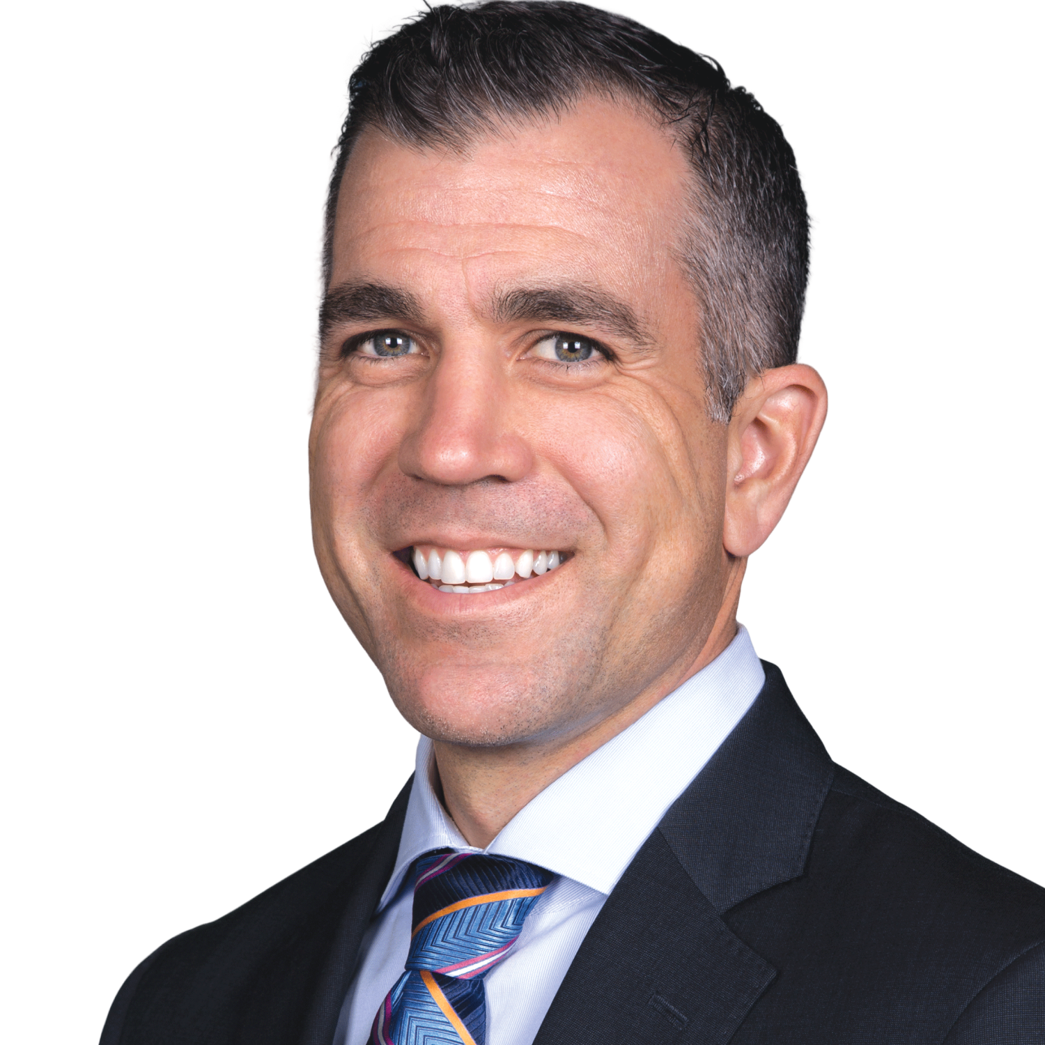
Dr. Duplantis has practiced for 24 years in Northeast Tarrant County, where he and Dr. Kirkham have partnered since 2003 in a cosmetic and restorative practice for patients of all ages. He completed the Advanced and Masters series at the Center for Aesthetic Restorative Dentistry in Dallas and later taught on its faculty. A consultant and key opinion leader for dental manufacturers, he lectures nationally on digital and CAD/CAM dentistry and restorative implant dentistry. He has been published in numerous journals and co-founded a social media group for dental education. He also volunteers with Save-a-Smile, Mission Arlington, Christina’s Smile, and White’s Chapel Dental Charity Clinic, which provide care to underprivileged patients.
Done learning and want to get started?
Book a demo