
Harnessing the Power of AI
For Better Precision, Efficiency and Patient Care
Transcript
Hello, everybody, and welcome to this webinar. Thank you for joining doctor Kunal Rai and myself, Rabiha. I am your host today. I am a dental hygienist and a customer success manager here at Pearl. We are going to talk you through how to harness the power of AI for better precision, efficiency, and patient care. So what I will do is pass it over to Kunal, who has kindly joined us. Thank you, Kunal, for joining me.
AI having me.
Hear what you're going to share. So I'll pass it over to you.
Okay. Good evening, guys. So my name is doctor Kunal Rai, and today, I'll be talking about harnessing the power of AI for better precision, efficiency, and patient care.
So in no particular order, I would like to give you an insight into Pearl second opinion, go through a few case examples of radiographic analysis, give you a quick tour of how second opinion works, and how second opinion has elevated my clinical and operational efficiency.
So I'll start off by introducing myself.
So my name is doctor Kunal Rai. In terms of education, I graduated from Charles University in Prague in twenty fifteen.
Since then, I've done a MSC in dental implantology with distinction from Manchester. I've done a PG diploma in restorative dentistry and a PG diploma in clear AI therapy.
To complement my dental implant work, I've done a PG certification in oral surgery.
I'm a general dental practitioner and practice owner, and I'll talk about my practice shortly.
When I'm not in practice, I run a one to one mentoring course helping fellow dentists with their clear aligner cases and to set it up and to get the best outcome for their patients. In terms of awards, I have won the ADA clinical case year of the year award in twenty twenty two, and I'm shortlisted finalist for the case of the year twenty twenty four, which I believe gets, we get told in in February this year about the winner. In terms of AI, I'm also a young dentist finalist in the year twenty twenty and twenty twenty one. Entrepreneurship, I'm a cofounder for a clear aligner system called AI.
I'm a key opinion leader for Tokiama and Sima Dental. In terms of charity, we donate a percentage of of our profit to Wells on Wheels as Shaz initiative, where I personally provide the kids with school uniform, books, and everything school related. In terms of social media, we do post cases online. My personal professional account is kunal AI underscore dentistry. And for the practice, it's meliora underscore dental.
Moving on.
So when I'm not in practice, this I'm basically working on focusing on the AI brand. I'm also a big advocate of digital and AI dentistry. The middle slide that you can see here, this is me talking at Leeds University about why we should work with digital digitally, why we should do digital dentistry.
The slide over here is me last year talking in November last year talking about AI, why, as a dentist, we should embrace it and not be afraid.
So the practice itself, the practice is called Meliora Dental. The word Meliora is a Latin word for above and beyond.
The practice I noted have been established for over twenty years and used to be trade under the name Roundy Road Dental Practice. And as you can tell from the name, it's located in Roundy Road, which is in Leeds, l s eight area.
The practice used to do the basic bread and butter of dentistry, doing a lot of assessments, a lot of silver fillings, and extractions with many plenty of plastic teeth. Over the last twenty four months, I basically I qualified in twenty fifteen, as mentioned, then I did my VT in Burlington.
And thereafter, I worked for seven years for Bupa. And in twenty twenty two, I decided that I'm gonna take the plunge and move to practice ownership.
That's when I signed up to Christie's Dental Aid, and I was introduced to Round the Road Dental Practice in May. I placed an offer in June twenty twenty two, which got accepted. And in February twenty twenty three is that's when we moved in, and this is what I acquired.
Black and white looking exterior and, bizarre looking black and white walls with some random red chairs, which we I can assure you we have got rid of. Over the last twenty four months, I have invested heavily into the practice so we can provide advanced treatment to our patient, improve patient journey, and communication.
We've also introduced various new tools, software, and one of the application that hell has helped me to move from hundred percent national health to a mixed practice has been Pearl. And I'll talk to you about how Pearl has helped me personally to enhance to advance my my practice.
So this is a practice today.
So we do a lot of cases, a lot of smile makeover.
And that's Lily who just finished her treatment, and we've just added thank you. We're giving her a flower and bunch of chocolates.
So this was the practice when I bought it, and this is the practice today.
Moving on. So an introduction to second opinion.
Second opinion is essentially an AI tool that has been developed by Pearl. Second opinion is the first clinical AI diagnostic tool that has been deemed safe and effective by the FDA FDA.
To my associates, to my staff, I say to use this tool as a second set of eye that can help with decision making and communication.
It is a real time radiographic analysis that can help to detect, which is certain anatomies, cavities, pathologies, bone loss, and so on.
So why did I choose AI radiographic tool? This all stems back to my time as a, as a VT train a trainer.
Base training. This basically so I was working in Burlington.
In second month of my training, I was I saw a patient over lunchtime, Mark, and I did his report, his and I basically was doing his report over lunchtime.
AI identified the upper AI four, upper eye five for a filling. And over lunchtime, doctor Mitesh who recently qualified came to my surgery to go for lunch. I started discussing this case with him, and he said, now I will do the upper right four as you are in five, but I also do the lower left seven d o because there was a bit of a shadow in there. Seeing us having a conversation, doctor Katrina, as it was lunchtime, doctor Katrina, a qualified dentist working in the practice for over five, seven years, she came to our room and we started having a chitchat and we are I asked her opinion on this x-ray. And she said, AI do the upper right four as you said, but I would do the lower right six. So in the span of twenty minutes, three dentists within the same practice gave three different treatment option for this same patient.
Subsequently, I spoke to my trainer doctor Karlie Cech and I asked for his advice and he he just said to the just just to do the upper AI four. So overall in a in a half a day span, I had four dentists, same practice, giving four different treatment plans. Now imagine if you were this patient and you went to see all four dentists, you would be damn confused who do I believe. They've all given me different treatment plans. So from that moment of time, I knew when the time comes and I have my own practice I will not have my patient walking out confused.
So AI the time came February twenty twenty three AI start this deal went through AI started looking into softwares AI started looking into tools and I had a demonstration for second opinion And from that moment of time, I was completely blown away with this software. I knew if I do not have this software in my practice, I will not be doing justice to my patient.
So I sat down with Jack. We discussed numbers. We agreed on a price, and the installation just literally took a few days. And it happened in the background with the IT team. Everything was done. Then Ravi AI came over lunchtime, virtually gave us a demonstration.
And since then, we have not looked back as a team, and we use this tool for both our NHS as well as private patients.
Okay.
So in a chronological order, I'm basically gonna talk you through what I've done so far.
So when I qualified, I basically used to spend a little bit of time using a magnification glass on the exact tools to look into teeth, see where caries are, and then I will present it to my patient. And this is what I would present. A black and white image talking about this is your this is your right side. This is your left side. These white blocks that you can see, these are your existing fillings.
This appearance is your bone. Can you see this little black shadow there? And nine out of ten times, patient would would not have an idea what we're talking about. They would just shake their head and either go to reception, book themselves for a filling, or just pay for the assessment or even not paying, just walk out AI never come back.
But one patient basically left as a Google review. He basically said, I came for a checkup, Went to see my dentist, a new dentist at the practice.
He did a assessment. He took some x rays and then said AI need to have three fillings. I didn't have any sensitivity, any pain, but he said I need three fillings. Stay away from this dentist. You're off and busted.
So we've had a good and bad I don't know about you guys, but what I've generally, when I just used black and white image in the daytime, in the morning time, I used to find it. I was fresh. It didn't take me so long. But in the afternoon, after lunch, I felt a bit tired, and it take me longer to identify things on x rays.
Then came COVID, and it was like a blessing in disguise for me. I didn't spend a lot of money going out. No holiday, so I saved up a bit. As an associate, I then invested into the iTero scanner.
So now I'm just gonna go through a chronological patient journey. So when I qualified, I used to spend a lot of time looking at a black and white image depending on the time of the day. If it was morning, relatively quick. But in the afternoon, I used to after lunch, especially, I used to feel a bit AI, fatigue kicked in, and I used to spend a lot of time on magnification glass making sure I'm not missing anything out.
And there was always I was always running late, and I used to used to show a black and white image to my patients.
So it's talking about this is your right side. This is your left side. Can you these white things that you can see, these are your existing fillings.
This is your bone structure. Can you see this little bit of shadow here? That's a cavity. And nine out of ten times, I would just have the patient nod their heads in the air.
That's fine. They would go to reception, book themselves in, not knowing what they're booking themselves in for, or they would go pay or not even pay, just walk out, but they will always leave a Google review. I had one patient who left us a Google review on the practice AI, on the plastic on the practice Google review. I came to see a new dentist at the practice.
He did the checkup. He took some X rays. On the X-ray, he told me that I need two or three fillings.
I have no pain. I have no sensitivity.
Just feels like he made it up. Stay away from this dentist, and that's AI I had.
Since then, I have then in twenty twenty, I invested COVID came. I invested in a iTero scanner.
As you know, we didn't go out. We didn't have any holidays, so I saved up a few bucks, and so I invested myself as an associate into an iTero scanner.
The primary purpose of this scanner was to do for Invisalign treatment. However, I also use it for my private patients so we could do a scan so we can amalgamate it with the x-ray so to give them a better understanding of what is happening with their teeth. Like in this case, this is the lower arch. We spoke about the patient needing a good clean because of tartar built around. Then we spoke about how there are silver filling. Would the patient like to have this removed for a white filling?
Thereafter, we also basically show the these patches over here and with the x-ray and said, these are teeth that need have got some cavities and may need some filling. So it was a better communication tool.
The biggest breakthrough k. And this was Chelsea, sorry, my nurse who used to do my scan for me in Pontefract.
She started off ten minutes per scan, and by the end, she was doing things under two minutes.
Then the breakthrough happened in twenty twenty three. As mentioned earlier, I bought my practice. I installed Pearl, and I no longer show a black and white image to my patient. This is what I show to my patient, and I'm just gonna run the video of my communication with a patient.
I recorded this over Christmas, so please ex excuse the Christmas outfit.
In this video, what I'm aiming to do is to give you an insight of how I explain my X rays to the patient, how I spend less time explaining the X-ray, and more time educating the patients.
So we took two x rays for yourself as part of the assessment, one on your right side and one on your left side. This is the black and AI to admit that in the past you would have seen, and I'm not sure if it made sense to you at all. So let's jazz things up. I use second opinion, which is a three d radiographic tool to help us explain your x-ray a bit better.
So I'm just gonna click on pathology, get that switched on, and two part. So this is your top right. This is your bottom right. What we can see here so if I give you a mirror right now and if you smile, you can see white surfaces. That white surface that you see is what we call the enamel. AI on an X-ray, this is your enamel surface, the outer shell.
Under the enamel, we've got some soft areas, which is the green zone. That is your dentin. Then you got your nerve, which is purple, which provides nutrients and blood supply to your teeth, and that's your root. Between two teeth, you got the honeycomb piece that you see. These are the bone structure that holds your teeth in place.
What we can see here is an existing filling that's on your back tooth. You have a tooth missing here.
Additionally, we can see some green squares. These are where tartar has built up, and I recommend a clean for your tea. AI this generally occurs due to not cleaning incidentally not using incidental brushes to clean in between your tea. Now we can see some pink areas. We can see some light pink areas and some dark pink areas or magenta areas. These light pink areas are what we call enamel caries.
With a good oral hygiene, with a good diet, you can stop this progression. And in some cases, you can even replenish the lost minerals.
However, when this pink area light pink area becomes a dark pink magenta area, that's when we recommend a filling. And as we can see here, this has progressed six percent into the green zone. This has progressed fourteen percent, and this has progressed seventy two percent.
So this one is quite deep. This potentially may need some invasive treatment.
Now let's look at your left side.
This is your top left side. This is your bottom left side. We've got existing fillings here. These are between your two premolars.
This is the pink areas. As mentioned, these are enamel caries. With a good oral hygiene, good diet, you can stop this progression and even replenish the lost minerals.
However, on your first big two on the lower left side, you've got a progression into the green zone becoming a dark magenta, which means I would rec like to recommend a filling here for yourself on this too.
You've got an existing filling here.
Next door to the existing filling on your second premolar, you've got a cavity developing, which is twenty percent into the green zone. So, again, a tube that I would recommend a filling.
All in all, you need one, two, three, four, five teeth that need a filling.
And as it came out of NHS patient, by default, these five teeth would be you will get a fillable filling and follow under a band two. However, you can always upgrade it and upgrade it to the FIPA AI, and that is a white filling.
And if this was AI my mouth, I would go for a white filling here because that's in my smile zone. That's again in my smile zone.
AI wouldn't mind a silver here, but it's completely your choice.
Again, I would go for a white and white here because, these are the teeth that people will see, and I was I would definitely like that to be covered up.
Okay. Let's move on. So now let's just, spend a few seconds on this X-ray. As clinicians, we can understand what is happening here, but as a patient, they will not.
So just to give you a bit of history, this was a NHS patient that came to see me with some excruciating pain on the top right side, which was keeping her awake at nighttime. Symptom wise, based on symptom based on the diagnosis, we identified this as acute apical periodontitis, but this is the x-ray that I showed the patient.
A picture of convulsion where and I spoke to a different part and explained to it my finding. So what I said to her that the problem that you're having is, can you see this inverted crown over here? That is the infection. That is what's giving you that pain. Now opt treatment option here are do nothing, a root canal, or an extraction.
Mind you, this patient was an NHS patient, and she wanted to save her t, and she chose a root canal. Thereafter, what I had, I had another conversation with her to explain to her this needs to be a private treatment. So what I did, I went over this premolar and I explained to her, can you see this purple part, which is the nerve? You can see the starting point and you can see the end, which is right at the end of the tube.
Whereas if we look here, we can see the starting point, but it stops halfway, which basically means that the end is not clear. It it needs some further work. It needs some extra tool for us to identify that path because it's close due to calcification.
Yes. We can still save the tube. However, I need to refer you to our specialist who can take care of this, which means that it will be a private treatment.
Are you happy with this?
Originally, initially, the patient was a bit unhappy, but then over AI, she understood and was and we've managed to convert it from a NHS treatment to a private treatment.
A full mouth X-ray, I normally take this for my clear line of cases or a patient where who comes in for a wisdom too. Again, a black and white image making no no all sense to patients. So I convert to jazz this up with some color information, and this is what I display to the patient.
This patient came to me with problems with the lower right wisdom two, and we can see clearly why. So I zoomed into it, and I explained to her. You've got AI wisdom too. Now, this x-ray itself is not it's just telling us it's very close to the nerve. And if we take this tooth out, it's gonna put a risk of damage permanent damage to your nerve. So I'll refer you for a CBCT so we can find out whether this tooth is in front, on, or behind this nerve, and that will dictate how we treat.
Okay. So now I would like to give you a quick tour of how second opinion would runs.
Hi, guys. So I'm just gonna give you a quick tour of the second opinion software.
When you log in in the Europe, you need to go on to second opinion dot hello pearl dot e u.
You will be presented with the home page displaying all the recent patient AI, which contain all the radiographic images that are automatically populated onto the site.
Now as you can see, the home page layout follows a simple and modern looking design with the most recent image set appearing in the top left AI, and all the sets are ring in a snake pattern down the page. Each entry displays basic patient information, including name, date of birth, and the number of images in the set.
At the moment, a notable feature, PHI protected health information is activated, which is located in the top right corner.
When activated, it replaces patient name with asterisk, providing a GDPR compliant view. I find this quite useful when showing the screen to the patients so you can hide the patient information, and you're not revealing anything.
If you need to, you can easily just hover over the asterisk, which can reveal the patient's name.
Now the system attempts to automatically mount images in the correct position within the full mount series.
However, as clinicians, we can easily drag and drop images to new position if adjustments are needed.
So if I click on rotate, this the image can be moved around.
Okay. Now whilst we are here, I'm also gonna show you a compare feature. So let's just go on this most recent AI wing.
Now I'm gonna click compare, and I'm gonna compare this March twenty seventh twenty twenty four X-ray with the seventh of July twenty twenty two. So you got X rays side by side, and you can basically track changes over time.
Just keep in mind, the effectiveness will depend on the consist consistency of the image captured.
K. So let's move on. Now before I forget, second opinion relies on images being captured through your regular imaging software and then synced to the platform.
As clinicians, we cannot simply upload an image into second opinion for it to read like any other AI software. And currently, it only works with two d images. Later on in the year, it will start reading three d images.
Okay.
Let's look at another set of x rays.
Now what I wanted to show you is how does second opinion report on certain conditions.
Let's look at bone measurements. This system provides automated measurements from the crest of the bone to the cement enamel junction, and they are color coded in white, yellow, and amber.
The rate for white is zero to zero to not two point five millimeter. Yellow is two point five to four millimeter, and amber is greater than four millimeter.
So this is amber sorry. This is yellow, so greater than two point five millimeter and less than four. Because this is less than two point five, this is showing in white. I find this feature quite helpful for periodontal assessment.
Okay. Now let's look at something else.
Let's have a look.
So this is what the purple square that you can see here, this indicates areas of notable margin, such as potential overhangs or open margin on existing restorations.
Now let's look at something else. And we see the green square here. This green square highlights area of radiographic calculus, which is useful to hygienists and dentists in treatment planning and patient education.
This is self explanatory. You can see there's an infection here. Let me put on the two part feature.
So any kind of periapical radiolucency will be outlined in blue.
Okay. Now let's talk about caries.
Now caries is is arguably the most well thought out feature. The system uses two shades of pink to indicate potential caries, light pink and dark magenta. The light pink indicates early caries lesion, insert the aries hundred percent into the enamel, which we which in my case, I'll generally just review. The darker magenta, which are caries which are extending into the dentin, and we need to restore it.
Another feature AI we can see is the percentage breakdown.
This system AI a percentage breakdown of enamel versus dentin involvement, which I will talk about later and how it generates revenue for my practice.
Hi, guys. So I'm just gonna give you a quick tour of the second opinion software.
When you log in in the Europe, you need to go on to second opinion dot hello pearl dot e u.
You will be presented with the home page displaying all the recent patient AI, which contain all the radiographic images that are automatically populated onto the site.
Now as you can see, the home page layout follows a simple and modern looking design with the most recent image set appearing in the top left AI, and all the sets are arranged in a snake pattern down the page. Each entry displays basic patient information, including name, date of birth, and the number of images in the set.
At the moment, a notable feature, PHI protected health information is activated, which is located in the top right corner.
When activated, it replaces patient name with asterisk, providing a GDPR compliant view. I find this quite useful when showing the screen to the patients so you can hide the patient information, and you're not revealing anything.
If you need to, you can easily just hover over the asterisk, which can reveal the patient's name.
Now the system attempts to automatically mount images in the correct position within the full mount series.
However, as clinicians, we can easily drag and drop images to new position if the judgments are needed.
So if I click on rotate, this the image can be moved around.
Okay. Now whilst we are here, I'm also gonna show you a compare feature. So let's just go on this most recent AI.
Now I'm gonna click compare, and I'm gonna compare this March twenty seventh twenty twenty four X-ray with the seventh of July twenty twenty two. So you go X rays side by side, and you can basically track changes over time.
Just keep in AI, the effectiveness will depend on the consist consistency of the image captured.
K. So let's move on.
Now before I forget, second opinion relies on images being captured through your regular imaging software and then synced to the platform.
As clinicians, we cannot simply upload an image into second opinion for it to read like any other AI software. And currently, it only works with two d images. Later on in the year, it will start reading three d images.
Okay.
Let's look at another set of experts.
Now what I wanted to show you is how does second opinion report on certain conditions.
Let's look at bone measurements. This system provides automated measurements from the crest of the bone to the cement enamel junction, and they are color coded in white, yellow, and amber.
The rate for white is zero zero to not two point five millimeter. Yellow is two point five to four millimeter, and amber is greater than four millimeter.
So this is amber sorry. This is yellow, so greater than two point five millimeter and less than four. Because this is less than two point five, this is showing in white. I find this feature quite helpful for pedantal assessment.
Okay. Now let's look at something else.
Let's have a look.
So this is what the purple square that you can see here, this indicates areas of notable margin, such as potential overhangs or open margin on existing restorations.
Now let's look at something else. And we see the green square here. This green square highlights area of radiographic calculus, which is useful to hygienists and dentists in treatment planning and patient education.
This is self explanatory. You can see there's an infection here. Let me put on the two part feature.
So any kind of periapical radiolucency will be outlined in blue.
Okay. Now let's talk about caries.
Now caries is is arguably the most well thought out feature. The system uses two shades of pink to indicate potential caries, light pink and dark magenta. The light pink indicates early caries lesion, incipient aries hundred percent into the enamel, which we which in my case, I'll generally just review. The darker magenta, which are caries which are extending into the dentin, and we need to restore it.
Another feature that we can see is the percentage breakdown.
This system AI a percentage breakdown of enamel versus dentin involvement, which I will talk about later and how it generates revenue for my practice.
I find these features very useful for patient education and deciding between preventive measure and restorative measure.
Okay. So let's move on to the final part, which I want to show you is how to do printouts.
So you can click here. You can select the x-ray that you want to print out, and then you can just click on print out. So, essentially, you can customize the print out option, which is helpful for patient handouts, referrals, or insurance claim.
When printing, you can select number of images per page, page orientation, specific image to include, and which pathologies to display.
So this gets downloaded as a p in a PDF format, identifying everything that we have discussed.
So tartar in green, progressed cavity in dark magenta.
In pink, we've got the disappeared cavity. Yellow, bone level. The white is enamel. Green is dentin. Purple is pulp. Blue is cementum, and yellow stroke orange, pink is restoration.
And you can select the right and the left side, and you can just print it off and provide it to the patient.
Right, guys. So I hope you found that video useful. Now I'm just gonna mention you, essentially, things that we can detect.
We can detect anatomical structures such as the ID nerve. We can check the bone level, measure bone, which is quite good for a periodontologist.
Then we can talk about root canals.
We can talk about fillings, whether it's metal or nonmetal, the type, the percentage breakdown, periwico radiolucency.
We can talk about cavities, light pink, dark pink, tartar biltura, measure root length, and any kind of marginal discrepancies.
So how has Pearl built trust?
Do thanks to sec Pearl, it's not, spending a lot of money on second opinion. The software is now able to detect small and big cavities with precision, and that helps us dentists to describe things with confidence to the patient.
Moving on, how has Pearl builds trust?
Thanks to Pearl in investing a lot of money in second opinion, their software is now able to detect large or small caries precisely.
And due to the precision, other dentists are able to diagnose things with confidence. And if patients sees confidence in their dentist, they are more likely to take up the recommended treatment.
Visual annotation.
We know a picture is worth thousand words. And as human, we believe in what we see rather than what we hear. And Pearl gives that clarity. It empowers patient to make the right decision about their dental health, empowers them to take up the recommended treatment plan. But above all, it gives them the understanding of why did the problem occur and what they can do in the future to prevent this from happening again.
Okay.
So how has Poe made an impact to the practice? As I mentioned, I bought my practice in February twenty twenty three. And for the last twenty four months, I have invested heavily into the practice to improve our the communication, our patient journey, and overall to give patient an advanced treatment and treatment that the patient can walk out with full confidence in them.
Since January last year, I have made it mandatory to my practitioner to all my practitioners, associate dentists, that they have to use Pearl, and we keep a spreadsheet for data collection.
What we have noticed is that since we have started doing the data collection, there has been a forty eight percent I AI, mandatory, we have seen a forty eight percent increase in the number of patients taking up one or more fillings.
And it's up to that forty eight percent, we have found that seventy five percent of these patients are now asking for an upgrade. AI they are moving away from an NHS filling and requesting a white filling, which automatically generates revenue for the practice. And one of the reason is the percentage breakdown.
So this is an X-ray of Otis, a patient that has been coming to the practice since he was since he was seven years old. So he's been coming to the practice for over twenty, thirty years. He is thirty seven, I believe now. He I saw him for the first time in June twenty twenty four. He came for a regular checkup.
IT guy by profession, just snacks usual three meals a day, just has a snack in between.
No problems. Not no pain from anywhere. Regular checkup. We did the medical history. We went through extra oral intraoral assessment, and we took some regular bite wings. On the X-ray, AI basically we showed him the colored version through second opinion, and this is what I explained to my usual way.
This is your right side. This is your left side. This is your enamel. The sort of greener and purple area are your dentin, and these are your nerve. These are your existing fillings. You've got some light pink hair, which is fine. With a good oral hygiene, with a good diet, you can stop its progression of the cavity.
But, unfortunately, you've got some areas which have grown into the green zone, and we recommend some filling. And this is the upper right five. You need a filling here, you need a filling here, and you need a filling here.
Then I hovered over the dark pink area and showed in the percentage breakdown. And if memory serves me right, this was about twenty nine percent. This was about fifteen. This was about if I see it was about twenty to thirty percent.
So I said to Otis, Otis, you are a NHS patient. By default, we will be giving you a silver filling under a band two.
Okay. You can either go for that or you can upgrade the treatment and you can go for a white filling. A white filling is v per item.
And Otis straightaway turned around and said, doctor Kunal, these are gonna be a massive silver filling in my mouth. I'm not happy to have them. I'm happy to go for a white filling. I already have a lot of silver, and because of my job and because of the amount of conversations I have with different clients, I would like to go for a white filling. So we sold something without really selling.
This was Maria. Maria is a mother of two. She was an accountant by profession initially. Now she's gone back to university, and she is a second year student law student.
She came to see me. She is a registered patient at the practice. She came to see me with a broken lower right six, which is this tooth. We went through the normal procedure, medical history check.
We went through extra oral. We did some intraoral assessment. Thereafter, we took some regular X rays. And on the X-ray, we found a few teeth that needed a filling.
And she self confessed that she has her oral AI. Her sugar intake has really changed over the time since she's gone back to university.
She doesn't really have time for herself. She's spending a lot of time studying and looking after the kids and the free time that she has, she's literally knackered and she just falls asleep.
So the oral hygiene has not been optimal.
We decided due to the financial constraint, we're gonna go for a hybrid model. We will do the back posterior teeth in silver and the teeth that come in the in a front, anteriors of her smile area, we're gonna do that in white. She also chose for this to be a ceramic private onlay.
So this is what we did for her. We did a silver filling here, and thereafter, we did some white filling in this area. This was the tooth of concern. We drilled it out, and then we built it back over tokiyaba, funny enough, and then we checked functionally to make sure that there was no occlusal trauma.
This is a radiographic image of Tamara, a junior doctor, Liverpool Hospital. She lives in Leeds. She came to see me because she was getting some symptoms for reversible pulpitis.
We took a PA for her. We showed her the color version, and we showed her the percentage breakdown, and it was seventy nine percent Seventy five percent. Sorry.
So I gave her the same options. She chose to go for a white filling.
This was the tooth of concern. We can see the restoration is AI is not optimal. There's some leaking restoration, and there's caries over here. We drill the tube out, and we can we can see that there's no nerve exposure. The removal is above approximately seventy five percent, so we can say it's relatively accurate, the percentage breakdown.
Then we built it back up again with Tokuyama and a view stain over there. And if I must say myself, the filling looks quite realistic, and we did check the bite to make sure there is no occlusal trauma.
Now, guys, don't panic. We are almost to the end. So why Pearl and AI?
AI in my opinion AI is is the is the few is the present and the future Generally when I speak to the dentist the main reason they hate to use AI is because they have this fear that AI is gonna replace them But luckily dentistry is one of those jobs which is safeguarded because AI can't do not have the emotional intelligence cannot show empathy. If anything it can make us dentists to perform better by identifying things at a very early stage. Using the radiographic analysis, we can say, okay. This is about twenty percent into the tooth. Would you like to have a filling? At the same time, it can also empower the patient to make the right decision about their health. It can give them a micro lesson in dentistry.
Now I'm just gonna go through a few testimonials between, from my patients, from my staff, and my associate dentist.
So this is Angela, a patient of the practice. She basically said I love the two part feature it makes it easy to understand what is exactly happening with my teeth.
Mariam whose x-ray I just showed you before and a treatment that we did she essentially said I can tell myself now where I need a filling just by looking at the X-ray. And she AI so pointed out at the left side herself.
Colin, bless him. He's been coming to the patient for a very, very long AI, even before I was born. And I started seeing him for February twenty twenty three.
When I showed him Pearl for the first time, this is what he said, and I quote, I have been coming to this practice for over ten years. Why has no one shown me my X-ray like this? I would have looked after it much better. Bless him.
AI, emergency patient that came to see me with a problem with his lower left seven, we took an X-ray, a PA for him, and we showed him the colored version. And this is what he said. I like the colored version of my X-ray as I can see exactly what you are talking about.
Moving on.
Now I'm gonna talk about my staff. So as I mentioned, I'm a big advocate of artificial intelligence and digital dentistry. Apart from Pearl, I also use Kiroku for my notes.
Currently, my nurse, Kaylee, she does all my notes for me. She just right. She didn't have to type anything. She just have to click buttons.
And since we install Pearl, she can just go on to search on Pearl. She can know exactly which teeth needs a filling, And she just does my AI, X-ray report for me too. But I just wanna make this out, clear from the outset that I still go through my notes. I still go through my X-ray to make sure she done everything correctly.
What she says is AI can now do your X-ray report. Go sit in the kitchen, have sipping onto your coffee and have a biscuit while I'll I'll do your report.
But other nurses says we seem to run on time now. And then I've got another one who said it makes it easy for us nurse to check beforehand which tooth we are working on in case we have not we have noted incorrectly on our list.
Moving on to dentists, doctor Namrata, an associate of the practice, basically, she said, I love the two part feature. It makes it easy to explain findings.
She also said we can present x-ray findings with confidence.
Doctor Aditi mentioned AI am no longer missing out things on x rays, and the comparison feature really helps patient to see the progression which can be a convincing factor in their care decision.
AI. So I'd like to open the door for questions. For those of you who would like to schedule a demo, I'm sure Robia will be happy to help you guys. Please scan on to this QR code, and then you can fill in your details.
Thank you so much, Kunal. That was really interesting.
I also wanna thank FMC for hosting us.
Of course, please get your phones out and scan the QR code to book your demos.
AI.
Hello.
Meet Your Host
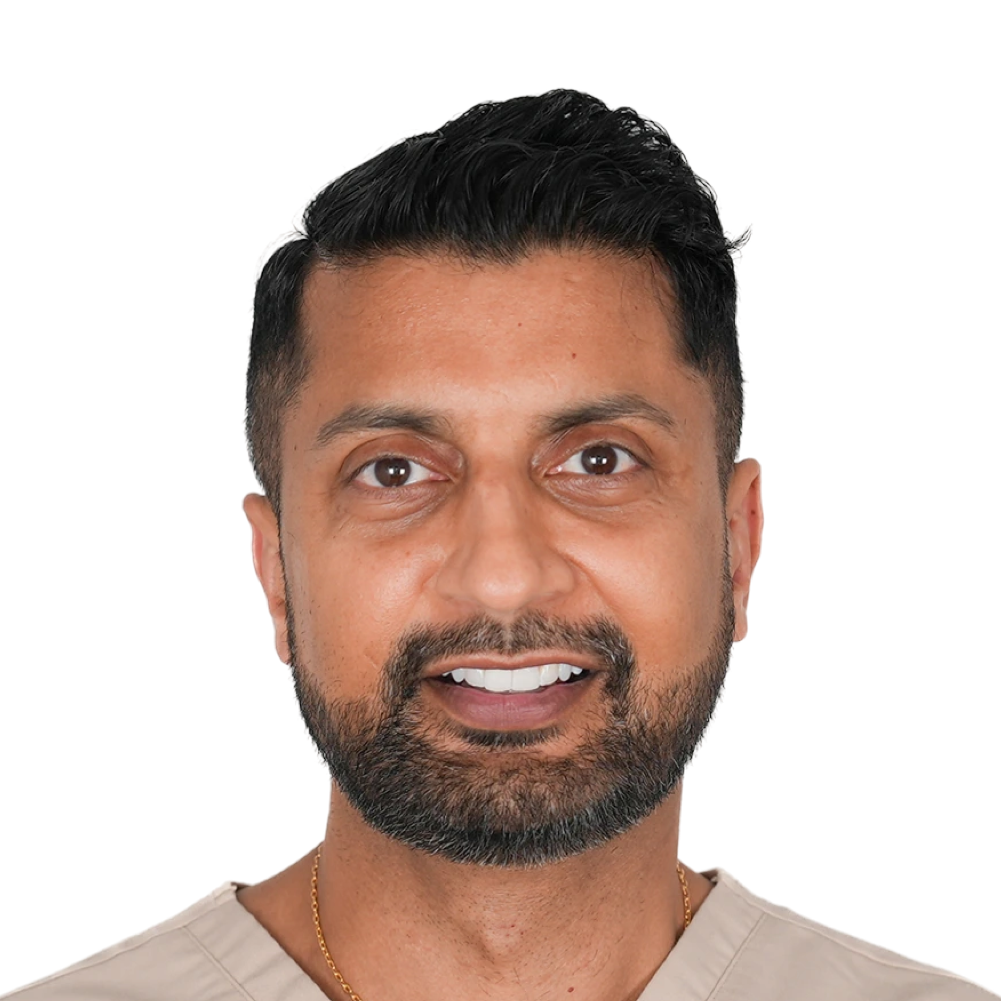
Dr. Kunal Rai graduated from Charles University, Prague in 2015 and has since expanded his expertise and depth of knowledge in dentistry. He has completed extensive postgraduate studies in clear aligner therapy and restorative dentistry; most recently obtaining a Master’s degree in Dental Implantology in 2021. Kunal is an advocate for Invisalign. He has a keen interest in cosmetic restorative dentistry, and is the most rewarding experience to seeing patient leave with a newfound confidence, achieved through mini smile makeovers which can range from orthodontic tooth alignment with Invisalign and teeth whitening to minimally invasive restorative treatments such as composite bonding, veneers, crowns implants and bridges.
Done learning and want to get started?
Book a demo