
Enhancing Treatment Presentations with Digital Technology
Transcript
AI of a fun topic, talking about really enhancing our treatment presentations with digital technology.
So with Pearl and Medit here, we're excited to have doctor Nezad walk us through what does this look like, you know, on a daily basis when we're interacting with our patients and taking that case presentation level, looking at it from a patient perspective. Right? Clinicians, we can look at cavities, from across the room, and we know if a patient's gonna need a root canal or if they need scaling and root planing. But really AI, we're gonna see what it looks like when we can include the patient, in their case accept a case presentation.
I am Sheela Roth. I am head of clinical education at Pearl. I am joined this evening with our, partner here, doctor Wong, who is also a general practitioner joining us from Australia.
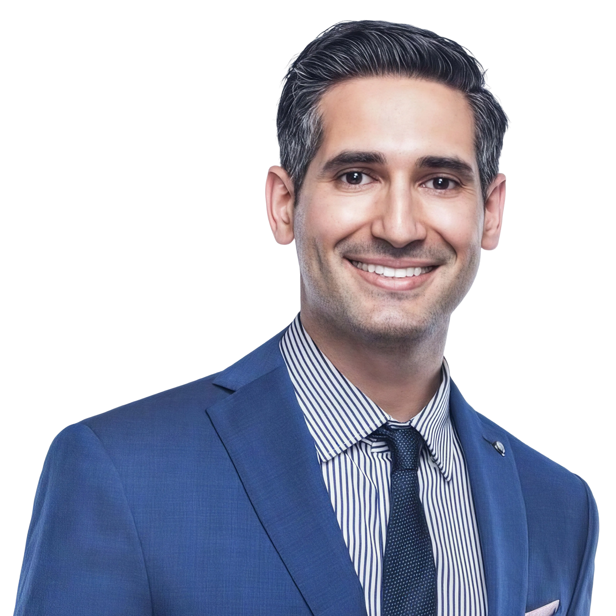
So we are happy to have Doctor. Najad here this evening as well. Doctor. Najad graduated from USC in twenty ten. He started his private practice in Beverly Hills, while at the same time teaching at USC as one of the youngest faculty members for twelve years. So, I don't know how you did both of those things.
So that's pretty impressive. He is a leader in the biomimetic and aesthetic dentistry realm, And he's also a fellow academy. He's teaching a lot in his own Nezhat Institute. We encourage you to visit that.
The other thing that I thought was really interesting, and I've also visited Doctor. Nezhat's practice, so I've seen this for myself, is that he really focuses on a patient centered approach and precision as well as quality over high volume. So, doctor Najad, thank you so much for joining us this evening. We're really excited to hear from you.
We'll turn it over to you and, again, encourage everyone to stay after so we can get someone on one time with doctor Nejad.
Okay. Thank you. First of all, thank you for that introduction.
Let me start my presentation here and share it. First of all, thank you all for joining. I'm very excited to share how I use digital technology to, elevate my quality, patient communication, all of the above. So let's go ahead and get started.
For those of you who don't know me, my areas of expertise are biomimetic and aesthetic dentistry. In my opinion, the two go hand in hand. But, you know, a lot of cosmetic and aesthetic dentistry doesn't look like natural teeth, and biomimetic is all about mimicking the natural properties of the teeth, restoring the integrity, minimally invasive restorations, and it it AI perfectly to front and back teeth. And as Sheela mentioned, I do online training.
I'll give you a little more information about that later. For today, I wanna show you how technology has weaved its way into my practice in a very big way. Okay? So let's start with digital technology and AI are reshaping modern dentistry, making treatment presentations more engaging and effective.
I have used digital dentistry for everything now over the past five, I would say actually seven or eight years now. But specifically in the way we communicate and share with patients, it's really made a huge impact on my practice. And it really just comes down to the power of visualizing and engagement. So patients need to be able to understand the treatment.
And it's just really important to realize that what we think they understand when we explain is, you know, a fraction of what they actually take away. So these types of tools provide better visualization, patients understand them, and it allows us a great way to communicate in a collaborative way with the patient. They start noticing what's this, what's that. When they come back in recall visits, I noticed them, picking out things, maybe getting concerned about something, and I can tell them if it's okay or not.
But that, both in AI radiographic diagnosis and in intraoral scanning has a tremendous impact on how we can share that. So take a look at this. You know, these digital tools here, the tooth parts, namely, which we'll talk about later, has a huge impact on how the patient can see, perceive, and understand what's going on. They don't understand when you point to the pulp area.
They need, they need some cues for these things, and this makes it easy for them to understand. Same with intraoral scans.
As just a simple demonstration of this, here is a full arch scan. And I was kinda just reviewing it in front of the patient, and you know what caught her attention? All this tartar here. So she saw it right away. I, you know, have her scheduled for a recall exam. It's not the first thing I'm talking about given her treatment plan, but she notices this right away. It opens up conversation and communication.
Another example, here is a case where I have upper temps. Okay? But look where I'm pointing right there. The temps have chipped in about a one month period. They should have last longer, but we noticed that these lower teeth are protruded and causing protrusive interferences.
I could actually take a scan of the, occlusion dynamic occlusion, the motion. But here I just zoomed in, showed her, AI to demonstrate in the mouth too. And what what this did was educate her, and the first thing she said is, okay. I need that Invisalign we talked about before. Because, you know, if the temps these are actually three d printed durable temps. It's kinda surprising, but you could see that when she was protruding, she was interfering on that spot.
As another example, this is called the occlusion analyzer. We'll get to that later, but this is the same case with an app that Medit has that allows me to look at the cross section and show her. You know what she said? She wanted her teeth to be, her final restorations to be less flared and less protruded. I use this cross section to show her in order to do that, one, we need to bring back those lower twenty four and twenty five. And, otherwise, you know, we're starting right up here at the gingival margin. I can't be too much more, constrictive.
I can't create less overjet than this and protect against even more chipping. So it sold itself for her to get the Invisalign and also was a great tool to, you know, effectively communicate the need for this, even though she was thinking about putting it off for a while.
We look at radiographs here. I mean, clearly, night and day, you can tell that from a patient perspective, it's a lot easier to understand this. You know? Try to show them the caries on number three mesial using the black and white radiograph, and you'll understand. And there's really no better way than doing an overlay. We'll talk about that in a little bit.
So in this webinar, I'm gonna showcase how Medit's intraoral scanning and the apps as well as Pearl's AI driven diagnostics can enhance treatment presentations, facilitate stronger case acceptance, and improve patient communication and outcomes.
So let's start with kind of the concept that digital tools really have had a dramatic impact in a lot of parts of dentistry, but very specifically in patient communication in this realm. It's I don't think scanning was made for patient communication, but it has that impact. And, of course, AI based radiographic diagnosis is made for us, but it has the huge benefit of also having an impact on our patients.
So one thing I'm an educator at heart. I've been told so many times by people that are not patients, but, like, let's say, staff members, other dentists, etcetera, that you explain too much to the patient. But that's what builds my patient trust, rapport. I like explaining, and patients tell me they love it.
And I think it's an important thing to spend time on. And traditional verbal ways take a lot longer. It leaves them confused. It's hard to verbally convey these things.
But seeing is believing. Since I started using intraoral scanning more routinely even in the treatment presentation aspects, it was a lot easier for me to convey these concepts, show them show them to the patient, and not have the patient looking in a mirror, squinting, and trying to see these things. So the power of identifying and seeing them large in front of them is huge, and it's also better in three d than it is in a two d format such as a photograph or a, intraoral photograph.
Like, intraoral camera, I meant.
I take really high quality DSLR photos, but I still reserve those more for before and afters, documentation. I find it's best to communicate these things efficiently with an intraoral scan and the radiographs that we need to get the job done. So digital tools provide clear visual evidence that supports the diagnostic findings.
So when we have these scans and AI driven imaging, we're able to highlight the areas. It removes ambiguity both for myself and the patient. I mean, there's definitely, we'll talk about this a little later, but there's definitely situations where I have had the AI be more accurate than I AI, and I'm very cautious in that. And that's the beauty of this.
It's not meant to, override or veto your opinion, but it's a second opinion. It gives you a little bit of information, makes you question things. It makes you also not miss things, and that's very important. From a patient perspective, it makes it very clear what they need, and that's a big thing.
So intraoral scanning and AI powered diagnostic enable real time discussions making, with the patient, making them more engaged in their oral health.
So this is a real clip from my, evaluation limited evaluation for this patient. They came in for number three. So I'm able to highlight these different things. I have a touch screen on the wall, really beautiful way to present these things, explain.
She had a temporary done by somebody else and, wanted to prevent a root canal. That's something I get a lot of referrals for with deep caries lesions. But I was also looking at her occlusion and noticing her teeth are worn. I can't improve the I can't really improve the anatomy of the tooth.
I have to go with the flat situation because I was able to use these tools to analyze that, and that can guide my preparation and my planning. So here I have those things. And also you'll see later AI set up for whenever we do restorative treatment. In this case, it was the same day.
So these tools streamline the workflows by reducing the need for repeated explanations.
They make it clear for the patient, and they improve overall patient acceptance of recommended treatments. I've had numerous times where the patient actually wanted the treatment based on something they saw that was inconsequential or not significant to me.
So in my routine evaluations of patients, they I've basically decided to incorporate both, Pearl, which is the second opinion diagnostic aid, and intraoral scanning routinely and systematically. So we're gonna go through both of these technologies in a little more detail about how they work and also how I'm using them. Okay? So first and foremost, let's start with the AI diagnostic aid.
So I consider myself an early adopter in AI, especially specifically within this program because the creator and founder of this program, was is a patient of ours at the practice. And one day he has, great experience and, previous success with a, computer vision related company related to advertising. And he said, what do you think if we started to bring computer vision to dentistry and use it for this? And we love the concept.
And early on, I did some annotation, some testing, helping kind of create the conceptual, components that would go into this. And I've been a user ever since. I've lost track, but I think we're talking, like, at least five, six years ago. Before this was on the market, we were contributing to its development, and it's amazing to see where it's come to now.
So, as an early adopter, I've got to see all iterations, help improve this, and I'm really, really happy with where this technology is right now. So Second Opinion is the name of the program, and it is FDA cleared real time AI diagnostic aid for dentists. Okay? It can do a variety of detections.
So this includes anything from bone levels, anatomical structures, segmentation of the tooth to include root dentin, pulp, etcetera, bone level, and even the progression of caries as well as identifying, you know, incipient and so on. We'll go into more of these, specifics as we move forward. And then it also has, image quality enhancement tools. You can work with contrast, clarity, etcetera, to help help visualize these things. And when the AI is looking at these things, it has the benefit that it's able to scan across a wide range of different views in order to form its opinion. So I find that if I'm editing the photo later to try to see these things, I can see it even if I haven't applied that particular, like, image enhancement with the utilities. But the AI, I believe, is using that in order to come up with its, suggestion or detection.
So we have all these different things. Periapical radiolucency is one of my favorites. I'll tell you more about that. Notable margins, great way to tell a patient about a defective restoration that could benefit from replacement.
It can identify restorations, fillings, calculus, cement that you leave behind. Residual cement will appear as calculus. So it's a great way to keep you honest and make sure that you go back and address those and so on. So it's a really cool software, that is making improvement all the AI, and there's so many benefits that we're gonna talk about right now. I really couldn't imagine, practicing without it now.
So normal image, we have on the left, pearl second opinion without the segmentation, just AI of the basic overlay including, like, you know, calculus, bone measurement, and, pathology or caries. And it's an interesting statistic that sixty four percent of patients don't understand the radiographs during consultations, but I can guarantee you they understand the pink highlights. In fact, there's a study that shows that it's better to highlight with an overlay to help educate a patient than it is to do a callout or an arrow. They still don't understand what the callout or arrow. You have to highlight it, box it. That's why it's like this. Highlighting is better than a point than pointing or an arrow callout.
When there's numerous things going on, I take this you know, if if these are the incipient AI, I take this opportunity to go over and teach patients that incipient lesions may never go away, but, will likely never go away. But if they're brushing and flossing and taking good care of things, they can avoid them from ever spreading. We can monitor that over time. There is a progression tool built in that will show you if something has progressed from previous radiographs.
So it's a really cool feature within there to help quantify and monitor things. That's also really useful.
There's different colors. So light pink would mean, like, an area where you're going to watch, whereas darker pink areas would indicate decay beyond the DEJ needing treatment. If you hover over this area, it shows you, like, the percentage within, dent within dentin that the caries has progressed. So it kinda gives you that quantifiable measure.
Periodontal images, also very beneficial and useful. So here we can see that there's definitely some probably moderate bone loss going on, some radiographic calculus, and look at these callouts. You know, we get the calculus.
This is really impactful for reinforcing the necessity for deep cleaning. I haven't found anybody questioning that. And I wanna reiterate, this is not about I don't think this is about patients trusting you or not. It's about them understanding and having peace of mind. And if they trust you, they will trust you. But nothing will impress them more and help them be more at ease than being able to also see it and have this validation.
So it's a very beneficial approach to planning. And it also honestly makes everything I do a little less stressful, a little more manageable. I enjoy having that second opinion to make sure I minimize misdiagnosis.
Tooth part segmentation. I thought this was super cool, but I didn't realize the impact. This is one of the coolest features that patients really love. You know, you try to explain periapical pathology, AI PDL, all these things, or try to explain that, you know, the caries here is approaching the pulp, but this is what helps them actually understand and visualize the problem.
Same thing with AI.
This is a really nice clean segmentation.
We can talk about the, incipient caries and the caries into dentin. We could talk about, in this case, you know, whether you treat or intervene. I personally would intervene at this point, but I would confirm with another radiograph, and then I would visualize the proximal surface of the adjacent AI nine percent chance there's no treatment there. But I explained that when I'm looking at one, we can look at the other. And AI, again, I use this as a as a way to show them the importance of flossing and determining if they're doing the types of preventative care that would avoid these things, and I have seen positive results from that.
Periapical radiolucencies are one of the things that are my favorite in here because I found that while this one is a very easy to spot periapical radiolucency, I've seen times where it has accurately diagnosed things that I am not so sure about. But given that we're gonna be doing treatment on the tooth, I, will either try to take an additional radiograph. Or if it's part of major treatment, maybe even have a consultation with CBCT.
And I find that more often than not, it's detecting things that I would have missed in this category. Periapical radiolucency is one area that it's significantly impacted my diagnoses and AI misdiagnosis.
I love that this engages my team. How many of okay. How many of you have staff that take a set of X rays and then come back, And you're like, I don't have this this contact. It's not open.
The purpose of the AI is to see the contact. Well, when they start using these tools, the segmentation and the, caries detection, I found them more actually absorbed into this process. So I'm finding that they're making improvements by being more aware and more connected to it and even being able to have discussions with the patients. So it gives them the opportunity to provide educational output.
It allows them to confirm what it is they're seeing. It's training for your staff, and it's an amazing thing to bring to the team, both for you, you know, your yourself, your staff, your assistants, even front desk, and hygienists.
Oops.
I don't really want this audio here. Let's see if I can mute that.
It's talking about how it may need a root canal. So, my favorite aspect of the software is this added educational value that I can just sit here, bring it up, use these different tools, and effectively communicate. It saves me time and elevates my quality. Patients appreciate that I'm an educator, and this tool has a big impact on that. It undoubtedly helps me and my team detect more and prevent missing AI.
That's, without a question.
And I love the patient printouts. I don't think enough people use this, but it reinforces the importance of radiographs.
We send it home with, patients after their hygiene visits when we take AI wings for any new findings or to reiterate what we have previously treatment planned.
So some statistics by the numbers. Forty three percent of caries and X rays are undiagnosed. Twenty percent of caries diagnosed in patients are not actual caries. Forty nine percent of periapical radiolucencies and x rays are undiagnosed.
I can say with the time and experience I've had with using second opinion, I personally fall into the category of making these same mistakes or contributing to these numbers. I definitely will miss caries occasionally, especially when I'm focused on something. I will definitely miss occasional periapical radiolucencies.
Sometimes it sees things that I just don't see, and I find that very beneficial.
And with this so a dentist that's using Pearl as a second opinion is thirty seven is gonna detect thirty seven percent more detections, and that basically means miss, prevent missing things. I wanna show you one way that actually, like, clicked with me right away. And I have, so let me just tell you the story. So here is post op X rays for four to thirteen veneers I did.
I'm just looking for cement. We got some cement between twelve and thirteen. I can clean that up. But when I take these X rays on the post op visit, I have tunnel vision on cement.
Okay? We're here to evaluate patients' inflammation or health, gingival health, I should say, and find the cement, remove it, check the occlusion. But I'm not even looking at the opposing AI. And, of course, that's not what should be happening.
But I have tunnel vision. I have a busy schedule, and I'm focused on the matter at hand. So having this visual cue automatically, reminds me to take a look at those things myself. So I'm just not even focused on it, and now I can see it.
And it leads to more treatment, but also prevents something getting out of hand and becoming a more serious problem. So I take this opportunity to review it. And if you have it in front of the patient, I can promise you they will bring it up and say, what's this? What's that?
So really great way to, remember the importance of looking at all those things. And if that doesn't apply to you, I wanna say it for sure applies to me on a routine basis.
The patient printout is a super cool feature as well. So here, what we do is we can select the images. This is great for AI wings, for example, and it can create this. I've blocked out the patient name.
So, basically, what we have here is a printout that shows the X rays you can do with or without the, tooth parts activated, and then you have the second page that shows the different findings. So it's a nice way to remind patients if they I I actually find that it minimizes them asking for a copy of their radiographs in the future. I think it minimizes the their need or their desire to get a second opinion because it's so clear and it's based on AI. So really cool feature.
And I was just told there's gonna be more development in this. I love the idea of incorporating this even on, new patients during their comp exam, just sending it like a template, and I wanna work with Pearl to accomplish that. But this type of patient report reinforces the value and the importance of prescribed diagnostic radiographs.
Patients are always trying to get out of them, trying to downplay their importance, trying to question why they need them. Things like this, I believe and have witnessed contribute to the value that is necessary for them to perceive that they wanna do this.
So that's a, a little intro or a pretty nice in-depth intro really into how I'm using second opinion. Now I wanna show you about intraoral scanning. So I think, many of us are familiar with intraoral scanning. It's not so much the technology itself. I think everyone is or a large percentage of people are already using it, but it's the concept that I have incorporated this into all my different comp exams, limited exams, consultations.
I used to take some photographs during the consultations, but I found that this was more valuable, and it has a lot of benefits for the treatment process.
So let's look at some of these things. So first of all, you'll notice my assistant is scanning. My scanning is included.
It does save me time overall and adds value. Saves me time in many different ways. I'll show you some of those ways.
So actually, I'll tell you some of them. One is education, other is for future appointments, if treatment's necessary, and even for emergencies, complications. Having a record is so useful. And it's so valuable that even without reimbursement, I would recommend it.
I incorporated it into my procedures without raising the fee for them. I felt like overall, it saves me AI. And my assistant has now gotten good at it, and it takes about three minutes, gives me something to work off of, gives a sense of high technology to the patient and has lots of utility for the future. And for everybody that says AI assistant, you know, like my assistant's not good at scanning or, you know, you people ask me, you delegate scanning to your assistant.
Well, I delegate it for these types of situations and I did it myself for restorative. Then as she got good, I let her do a large portion of it. I still take the final prep scan, but she'll do the pre op and all those things. And she's gotten tons of practice because we're doing it routinely.
If you don't do it enough, then they'll never get good at it. This is a perfect situation to allow your assistants to scan. And then when it comes time to review these things with the patient, it's such a useful tool for evaluating occlusion, teeth alignment.
You can even simulate straightening. We're not going into that though for this webinar.
Problems and defects from abfraction to recession.
We can, see chips and fractures, tooth wear, periodontal conditions AI black triangles, and even the tooth shade. I've had people literally look at it and go, oh my god. My teeth are that yellow. And it's it's funny because I'm not even really you know, it it does do a pretty decent job of simulating colors, but I don't think about it that way, but they do.
And it opens up these conversations. So great tool to incorporate for the reasons I've mentioned. And we can, like, zoom in close and look at all sorts of different situations. We have, first, we have a amalgam with fractured, enamel along the AI.
And then here we have a lot of erosion and acidity with failed composite restorations over the years. Here on this one, we have abfraction lesions and then, more or less the same here, some abfraction with a little bit of discolored dentin, maybe affected dentin in this process. So all good ways to communicate those things.
Here, I had a patient with a limited evaluation for treatment here. She came to me because she wanted to avoid doing a crown, and I'm known for doing, you know, biomimetic, minimally invasive. She wanted to save what little tooth was left there. So we took the you know, her focus was on number thirty. We took the radiograph, the PA, and the bitewing.
And then I realized, first of all, we actually let let's go back first. Actually, let's look right here. We realized that we should also potentially treat number thirty one because it also had some significant cracks, but my original PA radiograph did not catch the apex of number thirty one. So we go through our treatment.
We get to the end. And now because my team is so engaged in stuff, I literally was saved by my assistant. She said, should we take a post op PA, after the preps of number thirty one? Because it doesn't show in the original radiograph.
And I just totally had blanked on that because we had started the appointment focused on thirty and ultimately pivoted same appointment to do both teeth. So we took this PA radiograph, and lo and behold, we have a periapical pathology. We referred it for CBCT and treatment with our endodontist.
And, it was a huge periapical infection, really, really significant destruction of bone there with a very like, almost all buccal bone gone. But, yep, I can't say that I would have a hundred percent detected this if I was focused on the appointment with all the different things I have to manage. So I find this to be a very impactful part of my, quality control and assisting me in these things, and I have no shame in saying that.
It's about identifying how we can improve, and that's kind of the conclusion I've come to. Here's the final onlay restoration. We ended up temporizing number, thirty one so she can get a root canal. So we did a three d printed temporary there.
Here's another patient where it it highlights the impact of having good records. So because I had her record originally, I was able to explain this to her. So, basically, here we have her, scan from her comp exam, and you can see that the, restoration here is, it's, like, chipped and fractured, and I ended up repairing this for her at a subsequent visit. She didn't wanna replace it because it's a four unit bridge.
So we did a little composite there, and we replaced this. Okay? Then, she came back, and she said that, it chipped again, which, of course, is possible. And I said, okay.
It's, you know, probably time to do more treatment. But on closer inspection, I'm like, you know, it's not just that it has, fractured. It's also that more of the crown is fractured. So I'm able to use this tool.
It's Medit Design. I align the scan and the new scan. So now they're on top of each other. Very simple and effective way to do this.
And now you can make one of them transparent or play with the opacity. So look as I do that and zoom in, we can see that we have the, mesial buccal cusp has also lost more. So it wasn't just that area that was worn down that had been repaired. It was even further.
And this ultimately got her to say, okay. It's time we change that bridge. Isn't it? Said yes.
Thank you. So, here is here is the picture. So in twenty twenty two, then, a couple months a couple years later in, twenty twenty four. And, actually, she, is scheduled for tomorrow.
Now, intraoral scans also provide valuable records for tracking changes over time in other ways. So here, I have a patient that was referred to me because tooth number four has these notable margins, but also symptomatic. Okay? It doesn't look too bad in the scan, but radiographically, we have these notable margins, and she's symptomatic.
Okay? And, so after having a couple consultations, even with a root canal specialist who said, I don't think you need a root canal, but it's possible. I would try to avoid it. So she came to me at that point and said, you know, what do you think?
I did my testing. I said, sure. Yeah. I think we have a good chance. It tested with symptoms of reversible pulpitis, but it was vital, not necrotic.
We go in there, remove the restoration, use caries detecting dye, and, of course, there is leakage. This patient and this patient this photo isn't zoomed in enough, but you can see that there was a nice path of leakage from the mesial. And so, even through the temporary period, she said it feels better. Here is the final restoration cemented.
It's a Lissi inlay. But she came back.
She said that tooth is well, actually, she didn't say that tooth. She said that original problem is gone, but when I drink cold, I do feel sensitivity, and it's not going away. So most often, I find that to be cervical hypersensitivity.
So bring her back, take a look, and I end up blowing air on number, four, three, four, five, six, everything. And the only tooth that's sensitive is tooth number three cervical where we have this recession here. Now how did my record save me? She said, well, that wasn't there before.
And, so did you cause that recession? I'm like, hold on. Here's your scan from initial, which was, like, three or four months prior to treatment. Here's the scan of the day of, and here's your scan now.
It's always been there. She's like, oh, why is it sensitive now? So there's a lot of reasons. It could be bruxism.
It could be clenching. But the simple answer is you have abfraction. We can desensitize it. It may go away on its own.
But she was ready to blame me for causing something that I had nothing to do with, and it's my records that helped me save that, especially my initial because I'm able to, like, look at it and track it over time. Another super useful thing in my practice has been to have these intraoral scans. So I take them on the comp exam. And now when a patient comes in, in this case, the patient and I didn't do these, but, you know, one day one of them fractured.
And it's amazing to be able to replicate it and have that record and also to have something to temporize. So patient came in. I already had a putty created so I can temporize this. And, that is very beneficial.
And also, I wanna mention how beneficial it is when a patient calls in and has a problem. I've trained my staff to try to take a moment, try to assess where it is, even pull up the scan if necessary so we can schedule enough time to do the treatment. A lot of our patients want treatment at the same time, and I can't always promise that. But if we have a good understanding of what's going on and I feel pretty confident, we can make that sacrifice sometimes.
But it's these records that help us achieve that. And then one of my favorite tools from Medit is this, occlusion analyzer. So this is an app that allows you to look at cross sections and analyze the occlusion of the patient in great depth. So here I'm doing a composite restoration on the patient.
I wanna know if I have space to add any extra material there, because if I, you know, build it up and I put some anatomy and then there's tons of adjustment, that can be frustrating. So this app is really cool. I use it a lot. In another case AI here, the patient was saying that this crown she got, like, about a year ago feels high and it hurts when she bites on it.
So I can use this occlusion analyzer chairside, show it to her, and I saw I don't know if you saw it AI that, hyper occluded red contact spot showing her that it has a high contact spot, and we can adjust it.
But also it had more marginal integrity and an open margin. So overall, we lean towards, replacing this restoration.
This is the same patient as the case, immediately before this one. Just a quick video of how this occlusion analyzer works. So you've got this cross section. You can AI scrub through here and get to where you're gonna be. So I was gonna be restoring number thirteen here so I can even go there, take a measurement, and get a good understanding for what it is I'm going to how much space it is I'm gonna have to deal with.
So key takeaways from this webinar, traditional verbal explanations leave patients confused or uncertain about their treatment needs. Digital tools provide a clear visual evidence and a reference. It enables effective communication with patients. It supports diagnostic findings and your planned treatment plans or your planned treatments.
Intuarial scanning and AI powered diagnostics provide many tangible benefits, providing great value to your practice, and these tools improve the quality of care, your team's engagement, create treatment opportunities, and streamline communication and comprehensive care for your patients. It's a win on all fronts.
Did not want to do that.
Let's just go back a slide. So if you're interested in learning more about biometric dentistry, I just wanna mention AI also have online training, and it's, on the Nezhat Institute website. I got level one, level two. Lots of details on biometric dentistry and how to do restoration similar to our shared. And now I am finished, so thank you all for your attendance.
Excellent. Thank you so much, doctor Nejad.
So important welcome. All of this information.
I loved how you walked through everything and really AI made it very simple to understand. With all of us that are using digital technology, sometimes we think about how amazing and great it is. But, really, when we see the patient truly understanding, that's really the gift of technology.
So I thought it was interesting that you mentioned the sixty four percent of patients really have no idea what we're showing them on the radiographs. And I remember as a hygienist, I used to, like, you know, do the same thing, circle calculus or hold up pencils to talk about bone levels and such. But, really, this has become a way to kinda sit beside the patient and help them make more informed decisions.
I thought it was very interesting too, and it's probably very similar when you're using Pearl and Meta together, but you talked about how you save time. You don't have to repeat yourself during those case presentations. Can you kind of go into that a little bit more?
Yeah. So, like, you know, I'm trying to for example, I'm trying to explain overjet or where the the bottom tooth contacts the top tooth. And I'm trying to use my hands and make these gestures. I'm like, hold on a second.
I have a scan, and we can just bring it up right here. And I could show that to them. So it's just clearer and easier to explain because it's AI, you know, how everyone has those different models that they can use to explain everything from TMJ to implants? Well, it's like you have the most relevant educational tool, which is the patient's mouth and in a way that they can see right in front of you.
And you can bring it up and show it and highlight the problem. So I find that when I do it this way, I don't have to keep going over the same thing. And, you know, AI, a picture is worth a thousand words type thing, it sticks more. You know, it sticks more.
Even in future visits, it sticks. They come back for hygiene recall. I refuse to go into a hygiene appointment without Pearl Up because I need to be fast and effective. I don't have time to try to be AI this over here.
It needs to be up and ready to go because it'll save me time and make that hygiene appointment the amount of time it needs to be. Otherwise, I'm always going over.
Yes. Absolutely.
I time is so valuable, and I I just love that the patients are able to really, really get that and and be able to understand it. And you don't have to go back to the front desk and reexplain everything, right, when you're already moving to a different operatory.
Yep. So I do want to see if I could share my screen for just a moment. And, also, I want to, bring doctor Wong back on.
Doctor Wong is the clinical product strategy leader from Medit. And there was a question, doctor Wong, that was really AI of focused. I know you answered in in chat, but I think everybody would like to know the answer too is, how do you really keep the teeth dry while you're scanning? And so I'm sure doctor AI can answer that, but, also, if you wanted to, chime in there, that'd be great.
Sure.
To Oh, sorry. I was first.
You first.
Thank you, Matt, for sharing your great presentation, and Thank you.
It really speaks to our heart. Right? We love to do what we love, but we forget to speak the patient's languages. And so Mhmm. We we really, made that so clear for us as thank you.
I would love to hear your tips. I would love you to go first, actually.
So Okay.
So in terms of, teeth How do you keep the teeth dry when you scan?
Perfect.
Very simple for me. Obtragate, can't scan without an obtragate in the mouth.
And so we place the optrogate. We dry whichever arch we're gonna be scanning on. I find that you can more or less keep it dry for maybe one minute. So or dry enough.
And, you know, honestly, the Medit Scanner does excellent even with the, saliva and everything, but, of course, it's nicer to have that information interproximal and all those things. So what typically will happen is if it goes over about a minute of scanning, we'll just try one more time. It's also a good opportunity to stop the scan and assess areas you need to pick up. But very simple, just like that.
We don't do anything else. We don't have to rinse with anything or place, like, a suction in the mouth. We just use the obtrigate, keep the cheeks away. I do retract the tongue when I'm scanning the lowers with the mirror, and I hold with one hand, unless the patient's AI an angel and can keep their tongue away and keep everything dry.
But, typically, I'm gonna be retracting a little and or fighting the tongue to get the lingual surfaces.
AI, I am exactly the same. I use an optrogate, one time use for scanning and put it in the bin, a trash.
Yep.
The trash in.
One of the valuable things aside from every all the other things, you know, dry tips, suction, obtragate.
The medit scanner itself has a tiny fan that helps to defog the mirror. So that actually helps me, dry the tooth. Additionally, for upper back second molars, distal of the second molars, for example, one of the best tips that I've personally used is you actually have the patient turn towards you and and actually move the lower jaw to the AI. So that brings the ramus out a little bit and and get the patient to actually close a bit, move the jaw to the side like a pirate, and they really laugh and go along with you.
And I train my assistants to do the same so that they absolutely love that trick.
I use that one too.
And my assistants have seen it countless times, but I don't think they employ it. I'm gonna follow-up with them. But I usually will get you know, the first thing I do when I come in the room after these things is assess any areas that are missed and if it's impactful for what it is we're doing. And if it's a comp exam and it's just missing a little bit of pallet or something, I might let it go. But if it's an important area, something I wanna review, I go, and I always end up getting it. And even the smaller tips help for those areas too being able to I have the I nine hundred, so we can switch tip and use a smaller tip, and that helps as well sometimes. But always try to do everything with the large tip.
That's awesome.
Great.
Okay. So here's another question.
This one, I think I can answer.
There was a question that talks about we throw the terminology AI around pretty liberally, which is a really great point.
And the question was, what makes this AI a little bit different from the color enhancing tools that that we've been using for years?
And, really, I'm glad you asked that question because although AI is very simple and we see it everywhere, with Siri, Alexa, I mean, you name it, we're using it in places we're not even aware of.
But, really, AI goes beyond just the basic image enhancements.
It's a it's a process that go that we go through using a vast amount of data, and it learns to understand patterns and recognition.
And then what it's able to do is really focus on focusing on providing more insights and also mimicking what the humans do. Right? So like doctor Nejad was saying, in the beginning, you know, we taught the computers how to understand what a cavity was. And you can imagine what that's AI, in in real AI.
We did that in school, and it was very challenging. So teaching a computer, this is not cervical burnout. This is. This is a cavity.
This is not. And after lots of data, we ended up training a model, and Pearl's consistently working on that. So I I just wanna make sure that answer was really clear, because people do kinda think about AI as well. This is just, you know, different teeth colors.
And I wish it was that simple, but now we're able to really move into a space where we're seeing it in medicine. Right?
American Cancer Society talks a lot about how they're using it in breast cancer diagnostics. So I think AI is here to stay. The important part to, clarify tonight is that, like doctor Nezhat said, AI is a second opinion. I mean, Pearl is second opinion.
Right? The doctor is the first and final decision maker. So I think that's really important to make sure everybody's aware is we're not just looking at radiology and then having it diagnosed and doctor Nezhad can go to Hawaii all day. Right?
You need to be there to confirm the diagnosis. So, let's see. Couple other questions here.
Here's one for you, doctor Nezhad.
What do you do when a patient asks for a copy of all of the images? Like, every single image, every appointment, and not just a few selected ones. So that was an interesting question.
Oh, kill me. I hate that part.
Well, so when it comes first of all, let's talk about like, let's assume scans as well. The scan part, super easy because we can, use Medit's share feature, create an export, send them a link. Makes it so much easier and minimizes cluttering up emails with, like, larger files, etcetera. So I always use that feature to share that way and let them allow download access. You can set a password for it, but it's all through Medit Link. And so I use that.
You could also interestingly attach other things to it if you'd like. I've thought about that concept. You could attach the X rays all there and just send everything that way. But what we typically do is bundle everything up and send it with, Dropbox.
We create a transfer link, and, instead of saving it again to my emails, it's a transfer link. So it's kinda saved on Dropbox, but it sends them a link to download it. That works well for us, and we send it with our, email that's encrypted and HIPAA compliant. But I have thought about submitting things like this printout.
It's just that when they've officially asked for their records, notes, everything, you gotta give it all. Personally, I still never share an X-ray that is irrelevant. For example, a try in or something because I think it creates confusion. So when we need these X rays, and that's why I said kill me, it's because my staff can oftentimes put together everything.
And then I hate when someone sends me records that are just a complete mess as well, and I'm trying to sort through them. I don't want the try in that's not relevant or a picture of the temporary and get confused. You know? So I try to, like, spend the time to review them, but I let my staff prepare them first.
And we put them into Dropbox, and it is a time consuming process. And I've thought about it. You know, we never used to charge for that, but we've thought about doing it going forward because it does take some actual AI, and it is something that some of our patients just do routinely. And I could just be, build it into long established patient just wants a copy of their records.
But the patient that comes one time asks for that, and we have to spend, you know, some extra time preparing it. Sometimes I've thought about charging it, but I've only charged it a few times. We haven't done it systematically yet. It's just something we've been considering.
Excellent.
Yeah. I used to invite them back in and see if we could talk about it and pick up the charts and figure out what went wrong. Ninety percent of the time, it was a financial thing.
Yeah.
So a lot of AI, if we can get them back in the door, but, yeah, when they're like, I need everything, it's so consuming of our time. So I wanted to take a moment and, make sure I share my screen. I do want you to know from the pro side, there is a special offer today, and I have a really important question for you guys as well, doctors.
This is a great offer, guys, because you are able to try Pearl out, our second opinion feature, for free. So that's kind of a great thing to be a part of in during this webinar tonight.
When it comes to workflow change, I think there's a lot of concerns. Like, I have X rays, but I have my regular X rays, or how how do I incorporate scanners, and what does that appointment look like? Do we need to add ten minutes to each appointment?
Can you walk us through kind of how using technology actually saves time, which you've shown AI. But, really, when we're just looking at these things, maybe there's a barrier or concern that we have a lot of workflow change management tonight to deal with.
Yeah. You know, there is so if first thing that happens is if you're just implementing scanning, you need to I I firmly believe you need to be the expert of every technology in your office yourself. So you gotta spend the time to do it yourself. And, as an example, I've seen situations personally where other doctors who don't scan will expect their assistant to be able to do it and it creates frustration and it's kind of like finger pointing at that point. You have to understand how to do it when the time comes if if you wanna rely on it. And if your staff is trying to learn, you have to spend the time to train them.
So I found originally AI was doing it, so it was a little bit more of my time. So the workflow would mean that, like, I would go in there and scan them a few extra minutes, but then I would end up saving time on the explanation side. That's kind of the realization I came to. And originally, I was not doing it on every single appointment, but I kept doing I did it on comp exams as part of, like, you now establish.
But then I started to realize, you know what, even for a limited evaluation or a consultation, it creates so many opportunities to point things out, explain them. And even when they come back, let's say it's a limited evaluation for a crown, it saves me ten or fifteen minutes on the next visit if next visit if I already have it. So when you're balancing things between, like, saving time not doing it, but then on the next visit, you have to do it, I found that overall, it saves time because when you use it, you save time. When you educate with it, you save time.
And only time I would say you're not saving time is in the early process where you're trying to implement it. I would just put five or ten minutes extra. I mean, a long scan or really like, the x-ray part, first of all, is just the same. Right?
Like, the x rays the the way I talked about is just taking x rays. And then the scan part, very long, is ten minutes. You know, that's a huge big file that's bloated. But a typical scan is around three minutes for top and bottom total.
And in real AI, what's stopping, maybe five minutes, and my staff has gotten to the point that they can do it in between three to five minutes. And so it's worth it. It saves the time. It adds value to the appointment.
[continuing from the appointment-value discussion]
I take, and I saved those photographs for right before treatment. Like, AI instead of taking DSLR photos, which in my case show up on the screen right away or like in thirty seconds or a minute. But a lot of people, it's like you got to go take the flashcard.
It's really hard to set up that way. Well, here we scan. It's available right away and it has all those other tools for analysis. So it's just way more impactful, way more utility.
Excellent.
This has been such an informative webinar. Kimberly, I'm not sure if there's any additional questions you're seeing from the audience.
There's just one, Sheela, about, Pearl. And if you have the ability and, actually, it'd be interesting from doctor Najad to understand how often he's toggling between the Pearl highlighted view and then the original.
Like, how often do you go back and forth to see what Pearl sees and then remove it to re All the time.
Reorient yourself?
All the time. So, space bar turns it on and off. That's the keyboard shortcut I kindly follow it. So every AI. But I like to do that, and it's also a good way for me to, again, confirm, but also to catch things that I would miss. Sometimes, there was a period of time, I guess I should say now I don't do it this way, where I would go through, then I would toggle it on.
But now I like to go the other way around. I like to toggle it on, go through everything it finds, but also see if I find anything additional. And so I find overall, that's become my most reliable workflow, and it does save me time by kind of grabbing my attention. But, of course, I have to confirm everything myself.
Great. Yeah. Kimberly always tells a story about it being AI spell check. Right? When we're typing along and you get the little red underline and you're like, okay.
I'll go back to that in just a moment. And, it's a great reminder. Right? AI spell check's right and sometimes it's wrong.
But I think what's really interesting too is to leave you guys this evening with a interesting statistic.
I think it's also great to remind ourselves, like, why did we originally go into dentistry. Right? And it was to help patients. And we can help them make better decisions when we're using these types of technologies so that they are accepting the dentistry that you're recommending earlier and more often, which makes for a retained patient because it's less invasive and less expensive.
So we saw tonight we can save time, and we can really AI of put ourselves in in the patient's chair and see what they can see. Like, the patients are finally able to say, okay. I understand why I would need to get this cavity filled. Right?
So I I encourage you to reach out to Pearl and to Medit and think about ways to incorporate this digital technology into your practice.
I really wanna thank you again for joining us. Thank you to doctor Wong, and thank you to doctor Nejad for being here AI, and doctor Nejad, incredible presentation.
We look forward to having everybody back on our future pearl webinars, Matt. Great great night. Thanks. Bye.
Bye bye, everyone.
Meet Your Host
Dr. Matt Nejad is a leading biomimetic and esthetic dentist based in Beverly Hills, CA. A Fellow of the Academy of Biomimetic Dentistry, he mentors and trains dentists through the Nejad Institute and serves as a Scientific Advisor. He also contributes as the dental advisor to the Byrdie Medical Review Board. Dr. Nejad earned his DDS from the Herman Ostrow School of Dentistry at USC in 2010, excelling in advanced training for biomimetic and esthetic dentistry. After graduation, he opened his private practice and became the youngest faculty member at USC, teaching from 2010 to 2022. Known for his patient-centered, precision-driven care, Dr. Nejad prioritizes quality over high-volume treatment to ensure long-term success.
Done learning and want to get started?
Book a demo