
Clinical AI Roundtable
AI Conversations for the Entire Office
Transcript
Hello. Hello. Welcome in, and thank you so much for joining us today. We're very excited to have you.
As you can see, when you first join, we have a poll going. We'd love for you to say hello in the chat and see how many caries lesions you see in this image and drop your answer in the chat.
I told doctor Juan that I would put my answer in the chat, so I will put on my hygienist hat. And while we're waiting for everybody to wrap up with their patients and join us here on the webinar, I'm gonna take a look at this X-ray.
Thanks for joining us, everyone.
Don't be shy. This isn't a test, so there's no there's no right or wrong.
Okay. I feel like I'm back in hygiene school again looking at radiographs.
Welcome, attendees.
We're so happy to have you.
Right now, we are reviewing this radiograph to see how many caries lesions we all see.
I'll put my answer in. Let's see.
Yeah. And make sure to include, you know, everything you see. Even if they're incipient lesions that you normally wouldn't treat in your practice, you would just watch. Include those as well. Okay?
Okay. That changes my answer a bit. Let's see. Okay. There's my answer.
Okay. Alright. Seen a thirteen, a six. Okay. So we already have quite a range there. Ten, nine. Okay.
K. Well, doctor Juan, it looks like we're a couple minutes past the hour. So to be respectful of everybody's time, I'm gonna go ahead and kick us off. Again, thank you so much for joining myself, Katie Behl, and doctor Juan Castillo and our other Pearl team members at our clinical roundtable today.
I'm really excited for you to meet doctor Juan. He does an excellent job of bridging the gap of using Pearl and using it in the practice and what that means for our patients. Not only that, the foundation of AI. So with that being said, doctor Juan, why don't you kick us off?
Yeah. Thank you, Katie. So let's go ahead and and let's reveal how many caries lesions our our AI has actually detected. Okay?
So as you can see on the screen, this is what second opinion has detected on the bitewing. Okay? And according to the AI, it's detected eleven lesions in total. Right?
So let's talk about what just happened here. You know, some of you saw more than eleven lesions, and some of you saw less than eleven. So if you detected six lesions, then for you, the AI over called five lesions that weren't out there. So this is what you would call a false positive.
A false positive happens when the AI detects something that did not exist in your interpretation. It's very important that we understand it's an interpretation. So if you detected twelve lesions, let's say, then for you, the AI missed a lesion. And for you, that would be known as a false negative.
False negatives, obviously they happen when the AI does not detect something that it should have detected in your interpretation.
Here's the truth. Right? This is exactly what we'd expect. Right? X rays are all about interpretation and interpretations always leave room for a gray area of differing opinions, which is clearly on display here, given how much our interpretations vary.
So it's not that the AI got it right and we got it wrong. None of us can really claim to be correct. The fact remains that the only way to know which interpretations are a hundred percent right on a two dimensional image is to biopsy the tooth and confirm the caries lesion directly.
By way of introduction, my name is Juan Castillo, and I practice chairside for seventeen years.
I own two practices, and I spent several years working as an associate for a large DSO. I also worked as a dental claims reviewer for a major insurance payer. Today, work on the clinical AI side here at Pearl, which means that I spend most of my days thinking about exactly this problem. Right? Like how do we make sure that what's on the radiograph gets caught every time by every clinician regardless of how their day is going? I'm really excited about this session also because I think I hope this is gonna be the first of an ongoing series, so probably once a month or so.
The goal is to keep this feeling very collaborative and peer to peer, not a one way lecture. So as we go, if there are topics that you'd find helpful, things that you're running into chairside, I don't know, questions about how AI fits into your workflow, send them our way. This series should be shaped by things that are actually useful for you. Okay?
Today, we're gonna cover two things. First, how AI actually works and why would it sees might look different from what you see and what that actually means for your patients. And then second, we're gonna go ahead and discuss how to use a tool in second opinion called tooth parts, so that you can turn what AI finds into something your patient can actually see and color and act on it. Alright?
So let's go ahead and and dive into it.
If you're fairly new to AI, it's important to point out that AI is not a radiologist in some magical server room. Okay? People have so many misconceptions. It's not a second guesser, and it can't diagnose.
Okay? Very important. This cannot diagnose. It's it's you know? And it's also definitely not there to be a performance monitor.
Okay? How AI actually works, I wanna take a minute to explain the mechanism because I think once you understand how it works, everything else starts to make more sense.
So, to build an AI model like the one that Pearl uses for second opinion, you have to start with radiographs. I mean like hundreds of thousands of them. Right?
And you have to train calibrated dentists on how to annotate those images. They carefully look at each one and they say, okay, this is a caries lesion, this is a periapical radiolucency, this is calculus, and so on. Those annotations then become the ground truth. It's the labeled dataset that the model learns from.
Right? So the model then learns to recognize patterns in grayscale values of the images. So so a subtle shift in density signals demineralization. A halo around an apex suggests a periapical lesion.
So it's not reading rules. It's learning what disease looks like in pixels across an enormous range of image types, sensors, different angulations, different anatomies. By the time the model's deployed, it actually sees more radiograph than any dentist sees in a life lifetime, so it's it's it's pretty impressive.
Now that we have a basic understanding of how the model works and what AI can see, let's talk a little bit about what AI cannot see, right, what what it doesn't see. AI is another tool in our tool belt, but it's never gonna have what we have as clinicians. Right? As clinicians, we have a three d real time view of the entire oral cavity.
So we see things like bleeding, bleeding on probing, tissue texture inflammation. We can ask the patient if they have sensitivity to hot and cold, pain on palpation or percussion, all these kinds of things. So this clinical context is something that no radiograph will ever capture and it's the missing piece that AI will never have. Right?
So really you're talking about two kinds of vision on the same problem, you and the AI working together, catching more disease. The AI sees the radiographic patterns at a scale and consistency that no clinician can match across a full day. The AI doesn't become fatigued on any given Tuesday when you have, let's say, you have a difficult patient in room one, we've all been there. You have the lab calling you about a shade match, a patient in room three who doesn't get numb, and then you have in room five, you have that one patient that's interested in cosmetic dentistry and you'd like to spend some time on, but you're rushing.
Right? So the AI stays consistent, whether it's the first film of the day or the last film of the day.
At the same time, we as clinicians get to see the entire mouth with loads of context. So together, this establishes an elevated standard of care.
Now now that we've talked about how AI and and clinicians complement each other, right, let's get a little bit more precise about why the poll numbers at the beginning of the webinar buried a little bit. You know, this this variance comes down to two things, recall and precision. These are two terms that we use a lot in computer vision, right, doing what we're doing with the AI. In the opening poll, some of you saw as few as five lesions and others saw as many as thirteen.
Right? So the AI in this case detected eleven, however. So having a high recall means that the AI tries to detect every possible lesion in the bitewing. So it leans more toward the upper end, like the eleven that we saw in the opening poll.
The trade off with this is that it comes at the expense of precision. Right? So the more the AI detects, the more likely some of those detections are wrong.
That's what some of you will call false positives. We swing the other way and optimize for high precision, meaning that the AI only flags the lesions it's most confident about, then the number of detections drops maybe down closer to, let's say, six. You know? And then now you're gonna say that the AI is missing lesions, which we'd call false negatives.
So you have to think about this kind of like a tightrope or a seesaw. If we tune the model to be more sensitive so it catches more real disease, it starts flagging things that turn out to be nothing. If we tune it to be more conservative so every flag it makes is more trustworthy, then it starts missing more real disease along the way. There's no setting that is gonna eliminate both things, but we're constantly walking the line, you know, tuning toward the balance point that's more that's most useful for you.
You know? So the trade off, you know, when the model works alone is is what we just spoke about. Right? But it doesn't have to work alone, and and that changes the math entirely. You know? Research has shown that forty three this is really interesting. Research has shown that forty three percent of caries lesions that are visible on a radiograph go undetected by unaided clinicians, and nearly half of all periapical radiolucencies go undetected as well.
These aren't obscure findings in difficult anatomy. These are findings that are present in the images, and we simply are missing them.
Before you think that this is about skill, it's not. This is about volume, consistency, and the visual limits we have as human beings to sustain consistency across a full schedule of patients. It's not a skills it's not a skill gap. It's a human factors problem.
You know, I'd like to stop and kinda give you a, you know, a very relevant real life example.
I have a friend who's a great dentist, and he was generous enough to share this experience with me.
You know, less than a year ago, he had a patient who was getting married, and she wanted four veneers on her upper laterals and centrals. She was a patient of record for over five years, so it's not a patient that he was unfamiliar with. Prior to the prep appointment, they had done a mock up, she came in for a whitening appointment just to to prepare for for for the for the prep of the veneers. In the time between her last set of X rays and her prep appointment, you know, my colleague, he had implemented Pearl's second opinion.
So before prepping, he decided to take some X rays to update her record. And second opinion actually ended up flagging a periapical radiolucency on tooth number nine. So he hadn't caught it himself on prior films. The patient was asymptomatic.
The discoloration was not so noticeable, was minor, and honestly, was the kind of thing that it's easy to miss when nothing about the patient is telling you to look twice. Right?
So as you can imagine, it was a very uncomfortable conversation telling a bride to be who came in for four veneers that there's a bigger problem on tooth number nine.
But it also meant that he could address it now on his own terms instead of finding out mid prep or worse, you know, cementing the four veneers over a tooth with a periapical lesion. So that's the difference between a hard conversation and a genuine crisis. Right? So interestingly, when AI and clinicians work side by side, thirty seven percent more disease is detected.
So this is a huge benefit for our patients and a safety net for us. And there's a very practical side to all of this as well. When we pair AI with with our clinical findings, it doesn't just catch more, it actually speeds up your read. So you're not staring at every surface on every film wondering if you miss something.
The pattern layer is already flagged for you. So your exam time goes toward confirming and deciding, not towards searching. Right? So it's it's time back in your day and not just accuracy gained.
One final thought I'll mention about your partnership with AI is that the clinician is always in control. K? This is another thing that's very important. It's you have to think about it kinda like spell check.
Right? Spell check underlines a word. It thinks might be wrong, but it doesn't retype your sentence for you. You still decide if that was a typo or just a name it doesn't recognize.
Same thing here. Second opinion flags a finding and you decide to delete it or keep it and what it means for your patient. It truly is a second set of eyes and having the second set of eyes is half the story. What you do with that finding and how you communicate it to the patient sitting in a chair is where the real magic is going to happen.
That's exactly where one of my favorite tools, inside second opinion comes in. It really does make a huge difference in treatment presentation and case acceptance.
But before I show it to you, I want to walk through three challenges we all face as clinicians that get in the way of communicating effectively with our patients. I think it's something that you're all going to be able to relate to.
So let's think a little bit about how long it took us to learn to read a radiograph. And I don't mean just pass a test on it, but actually get comfortable looking at a bitewing and knowing what you're seeing. It took years. Right? It's a skill we learned overnight.
So think about what we do with that skill. Right? We pull up a black and white image on a screen. We point out a gray shadow, we say, you see that right there?
That's decay. You know? We need to restore it. And the patient looks at the screen.
He nods. Yeah. You know, I see it. You know? I see it, doc. But the truth is most patients have a hard time understanding what we're talking about on black and white films.
Very few know what they're actually looking at, but they just nod and carry on. Many times they leave without scheduling treatment, not because they decided against the treatment, but because they walked out of the room not really understanding what was wrong or why it mattered. Right? So there's also the reality of our profession's reputation.
It's unfortunate, but I'll say it plainly. Dentistry has a trust problem. We didn't earn it yesterday and it's not fair, but it's real. Patients come in a little guarded.
They've heard stories. They've had bad experiences, so the wall goes up really fast.
Especially, think about when you have to walk in and you have to tell them that they need treatment on a tooth that doesn't hurt.
That's a difficult task. Think about it from where they're sitting. You're asking them to spend money, sometimes significant amounts of money on a problem that they can't feel based on an image they can't read from a profession they've been told to be skeptical of. So this is this is really a difficult task, and and it's something we have to do every day. Right? So you can be the most ethical, most skilled clinician in the room, and you're still gonna lose the patient's trust. Not because of anything you did wrong, but because you couldn't make the invisible visible to them.
Right? And then there's the downstream constantly affects the patient. When we fail at the presentation moment, the patient declines treatment. And that decline in treatment doesn't erase the disease, it just delays it.
So this isn't just like a production issue, it's a patient outcomes issue as well. The treatment they declined today is the emergency they're calling about in pain eighteen months from now. So again, things we have working against us, the years it takes to read a film, the trust we haven't earned yet, and the disease that doesn't wait around while the patient decides. You know, these are three real problems.
And there's one tool inside second opinion that goes after all three of these at once, and it's called tooth parts.
Know, parts, it it really allows us to seek speak to our patients in terms of color and not just in shades of gray. You know, this is now night and day in terms of communication.
In this slide that you're seeing now, we're looking at caries detections as they normally appear in second opinion. Most of you are probably viewing your X rays with the overlays detecting incipient and progressed lesions as you see here on the slide, And this should be part of your normal workflow. Okay?
If you're not familiar with tooth parts or haven't used it yet, you can simply turn it on by clicking over the molar icon in the toolbar labeled tooth parts. Okay? It's it's located right in this area here. If you go down to the toolbar, it's a little icon that looks like a molar. It says tooth parts next to it next to it. So that's you just click on that, and then you'll get the the the different colors and the different layers of the tooth.
Before tooth parts, we would look at this black and white image that you see here, we tell our patients, you see this dark gray area here on tooth number thirteen? This is a cavity, and it's super close to the nerve, which is the dark gray area in the center of the tooth.
Once that cavity expands and reaches the nerve, you're going to feel it and you're going need a root canal. Meanwhile, this patient has no pain. They're squinting at the gray shadows trying to take your word for it, and then they tell you the famous words, Doc, I'm only here for a cleaning. I have no pain. You know? So the skepticism is really building up quickly.
Now, you know, what if we pulled up the same image with tooth parts? Instantly, it comes to life. Right? I mean, now you can say, you see this magenta area here, this circle?
That's a large cavity. Once it touches the purple area in the center, that's the nerve. You're looking at serious pain, a root canal, and a crown, right? These things always seem to blow up the day before vacation or the night before Thanksgiving.
It never fails, right?
So let's treat it now and skip the pain and the bigger bill later. It's a completely different conversation that you're having with your patient, and the tool's really doing all the heavy lifting for you.
Now I can tell you that toward the end of my chairside career, I was able to implement second opinion in my practice.
My only regret truly was not having it during my earlier years.
My workflow was simple. I'd review the detections. I'd confirm they aligned with my own clinical interpretation based on my clinical context. Then I'd turn tooth parts on and I'd leave it on for the rest of the visit.
There's a reason for that. If I stepped out to see another patient or grab a staff member, patients would turn to whoever was in the room and ask, What do you think? I really need that feeling? I can see it now, but I want to just confirm what you think.
In that moment, your team can confirm the recommendation instantly because the color makes it easy for everyone, not just a doctor to communicate the same finding.
So it worked the same way for my hygiene recall. The moment my hygienist took x rays, she'd leave tooth parts on and it would spark a conversation on its own. By the time I walked in, the patient was already anticipating findings and ready for the treatment discussion.
You know, tooth parts really does make the invisible visible. When patients can actually see what's wrong, something fundamental shifts in the room. The conversation stops being your word against their skepticism. It becomes, here's the evidence, here's what it means, and here's what we do about it.
So the moment you can show a patient the exact surface you're talking about in color instead of describing it, your case acceptance improves. Not because you get better at selling, but because you get better at showing.
It also just it makes you more efficient at the same time. Instead of hunting for the right words to describe a shadow on a screen, you point to the labeled surface and the patient already sees it. You know? That that case presentation that used to take you maybe five minutes now takes you one minute. So you get the acceptance and you get the time back. Right? So I think those are great benefits.
You know, patients get the treatment they actually need. It's they get it. It's earlier treatment, simpler treatment, less invasive treatment instead of the treatment they end up needing because they waited too long. So it's better for them. And, you know, downstream, it's better for your production as well. Right?
So before I wrap up, whoever you are on this call, I know you're carrying your own version of a hard position. Maybe tech gets handed to you with no say in the matter. Maybe every tool, every dollar is a decision you own alone. I've sat in both chairs as an associate and as a practice owner and now I'm on the AI side, so I get it.
But there's good news. We're all solving the same problem. Not enough hours in the day and trust that has to be earned one appointment at a time. Right?
So that's exactly what second opinion and tooth parts are built for. The you know, not another tool pushed on you. It's actually real value for you and your patients. So, you know, when a patient sees what you see in color, it's not a sales tactic.
It's it's trust built in real time, and that's really what we're all here for.
So that's my presentation. I really appreciate you all taking the time to meet today. Next time we got together, I thought that maybe we could you know, go a little deeper on on possibly using annotations to improve insurance claims, you know, the outcomes. I think that would be really cool. I think that would be a great session. But we're definitely you know, we're open to to suggestions. Like I said, I want this to be very, like, collaborative and peer to peer.
You know? So let's go ahead. We'll open it up for some questions. I've got a few minutes left. So if you have any questions, please go ahead. I'm also going to move the slide so that you can see the QR code so that you can obtain your your CE. Okay?
Well, thank you, doctor Juan. I think that was a really interesting conversation, especially the beginning half for me. When I first joined Pearl, I had this assumption that AI was this magic wand. That once they implemented AI, it was going to solve, you know, all of my problems and see every single thing that I have missed.
But it's really important, I think, that recall and precision and understanding clinical context that while the AI is incredibly helpful, we'll never be able to operate on its own, and that's why it's so important that dentists are using their clinical judgment alongside the AI. Right? We're all better together. And, really, that clinical context is important.
The AI is only seeing the X-ray. It doesn't know the patient's age or the demographic or what their mouth looks like intraorally, And so that context matters alongside the AI. And a final note for me and from my hygienist lens is I really liked tooth parts and the colors even for preventative care. I I think a lot of times we get AI with the assumption that it's here to treatment plan and diagnose and do more restorations.
But in my mind as a hygienist, I really see it as a preventative tool. I can now show my patients much more easily those early incipient lesions in the enamel rather than squinting our eyes at it. So if we're using products like Curodont or I need to show them where they need to floss a bit better. You can also use those colors for preventative conversations as well.
Then those bone levels being a roadmap for active bleeding and evaluating for SRP.
I think the most important part in the workflow is probably that hygiene exam and having the X rays pulled up on the screen ready to go for your doctor because that's where we're doing that evaluation.
That being said, keep adding any questions to the chat. There is no right or wrong questions. I love questions. It gets the conversation going. So please don't be shy, and don't forget to grab this QR code so that you can receive your half credit CE for joining us today.
And please make a note of doctor Juan's email. We'd love to hear from you through our contact us form on our website.
Did you like this webinar? What other topics do you wanna see? Is there any feature requests that you would love to see in Pearl? We always love to hear from you and see what we can add to our product to better suit your workflow.
Any final thoughts, doctor Juan, as we wrap this up?
No. I mean, I I think I'm I'm excited about this. I think this will be great because, you know, hopefully, we get some suggestions as to what, you know, clinicians, really would like to talk about and discuss.
Because, like I said, I think, you know, the the clinicians are are the boots on the ground. Right? So it's important to to see, you know, how they're they're feeling about the, the implementation of this product and and and, you know, what we could do to make it more useful and for them to really understand the value in it.
Yep. Exactly.
Okay.
Well, if we don't have any questions, we can go ahead and give you four minutes back of your time so that you can go get settled in before your next patient arrives. I hope you found this webinar helpful. We hope to put it on again for you all, covering different topics and really ensuring that you're getting the most out of Pearl and connecting with other clinicians who are also using the product.
I hope you all have a fantastic rest of your week, and I hope to hear from you soon. Thanks, doctor Juan.
No. Thank you, guys. Appreciate it. Bye bye.
Meet Your Host
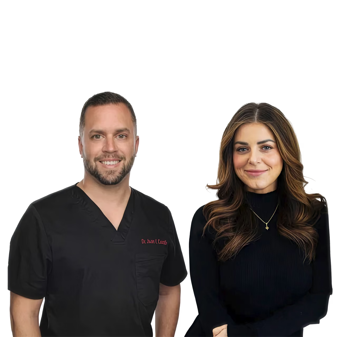
Dr. Juan I Castillo is Lead Dental AI Specialist at Pearl AI, working at the intersection of clinical dentistry and AI. A licensed DDS with nearly two decades of experience, his background includes solo practice ownership, three years as a DSO associate, founding two practices, and dental claims review. These varied perspectives shape his view on where AI can genuinely support clinical judgment. Dr. Juan brings an authentic practitioner's voice to conversations about AI in dentistry. Q&A by Katie Bayle.
Done learning and want to get started?
Book a demo