Dental X-rays are one of the most important tools your dentist uses to keep your mouth healthy. These images allow dentists to see problems that aren’t visible during a routine exam, including cavities between teeth, infections below the gumline, and even early signs of bone loss. In short, dental X-rays help catch small issues before they turn into major problems.
Understanding how dental X-rays work, what types exist, and when they’re used can help you feel more comfortable and confident during your next visit. Today’s technology uses incredibly low doses of radiation, giving your dentist clear insight while keeping you safe. Whether you’re wondering if dental X-rays are safe, how often you need them, or what to expect during the process, this guide breaks it all down.
What are dental X-rays and how do they work?
Dental X-rays (also called dental radiographs) use a small amount of radiation to create detailed images of your teeth, jawbones, and surrounding tissues. These images reveal things that can’t be seen during a visual exam, like cavities forming between teeth, hidden infections, or early signs of gum disease.
The process is simple. X-ray beams are directed through your mouth and pass through your teeth and bones at different rates depending on the density of the tissue. When they hit a sensor or film inside your mouth, they create an image. Dense areas like fillings or enamel appear white, softer areas like gums or decay appear darker, and everything in between shows up in shades of gray. This contrast is what allows dentists to identify everything from deep cavities to problems with root structure or bone levels.
Types of dental X-rays and what they show
Different types of dental X-rays are used depending on what your dentist needs to look at. Each one gives a specific view of your mouth and serves a distinct purpose in diagnosis or treatment planning.
Bitewing X-rays
Bitewing X-rays show the upper and lower back teeth in a single image. They’re great for spotting cavities between teeth, especially the spots that toothbrushes can’t reach, and checking the height of the bone between teeth (called the alveolar crest). These are commonly used during checkups.
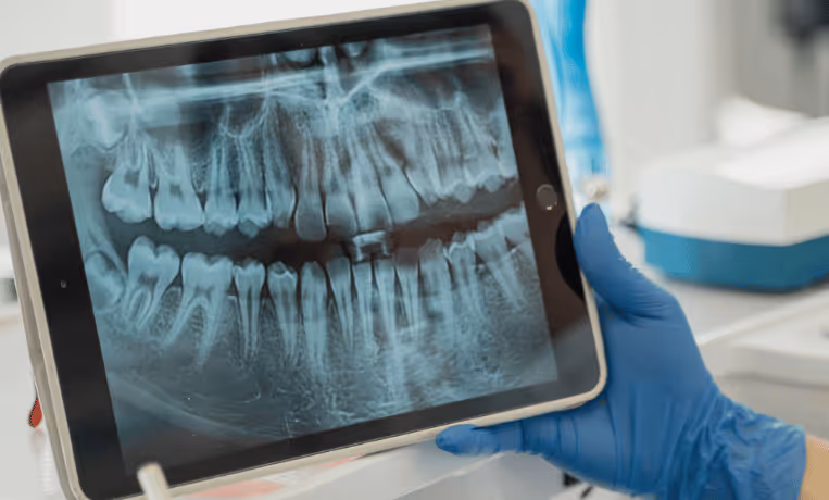
Panoramic X-rays
Panoramic X-rays take a full image of your entire mouth, including both jaws, all your teeth, your jaw joints, and even some sinuses. This type of X-ray is helpful when your dentist needs a big-picture view, like during orthodontic planning, checking for impacted teeth, or evaluating jaw development.
Periapical X-rays
These X-rays focus on just one or two teeth at a time, showing everything from the crown to the tip of the root and the bone around it. They’re used when your dentist suspects issues like infections at the root tip or needs to monitor healing after treatment.
When do dentists use X-rays?
Dental X-rays aren’t just for checkups. They’re used throughout many different parts of care to make sure your diagnosis is accurate and your treatment is safe and effective.
New-patient exams
If it’s your first time at a new dental office, your dentist may take a full set of X-rays to get a baseline of your oral health. This helps identify any hidden issues and gives a complete picture of what’s going on in your mouth.
Finding hidden cavities
Some cavities start between teeth, where they’re impossible to see with the naked eye. Bitewing X-rays are designed to catch decay early, before it becomes painful or expensive to fix.
Tracking child tooth development
Dental X-rays help monitor how teeth are growing in children and teens, whether there’s enough space for permanent teeth, and whether orthodontic treatment might be needed. They’re a key part of ensuring development stays on track.
Planning dental treatments
Before you get a dental implant, braces, or a tooth pulled, your dentist will use X-rays to understand the exact shape, size, and position of your teeth and bones. This makes procedures safer and more predictable.
Checking for infections or bone loss
Periapical X-rays help detect signs of infection or inflammation that might not cause pain right away. They’re also used to check for bone loss, which can be an early warning sign of gum disease.
Are dental X-rays safe?
Dental X-rays use very low levels of radiation. In fact, a set of four bitewing X-rays typically exposes you to less radiation than a short flight on an airplane. Thanks to advances in digital imaging and equipment design, modern dental X-rays are safer than ever.
Dentists follow strict safety guidelines. Digital sensors reduce exposure, high-speed settings minimize scan time, and lead aprons with thyroid collars can be used to protect sensitive areas. The technology is designed to capture clear images while keeping your exposure as low as possible. In most cases, the benefit of early diagnosis and prevention far outweighs the tiny amount of radiation involved.
Are dental X-rays safe during pregnancy?
Dental X-rays are considered safe during pregnancy. According to the American College of Obstetricians and Gynecologists and the American Dental Association, essential dental imaging can be performed during any trimester if there’s a clear clinical need.
That said, elective X-rays are usually postponed until after delivery, just to be cautious. But if you’re dealing with dental pain or a suspected infection, X-rays are often the safest path forward for both you and your baby. Untreated oral infections can carry more risk than the tiny amount of radiation from a properly shielded dental X-ray.
What to expect during a dental X-ray
If you’ve never had dental X-rays or it’s been a while, here’s what the process usually looks like. It’s fast, painless, and designed with your safety and comfort in mind.
Protective apron
Before the X-ray, your dental team might place a lead or lead-free apron over your chest and stomach. A small collar might also be used to shield your thyroid gland. These protective barriers help block any scatter radiation from reaching the rest of your body.
Sensor placed in the mouth
Depending on the type of X-ray being taken, you’ll gently bite down on a small digital sensor or film holder. Your dentist or assistant will guide you so the sensor is in just the right spot to capture the image needed. It might feel a little bulky, but it’s quick and should not be painful.
The machine is aligned for accuracy
The X-ray machine will be positioned near your cheek or jaw. The tube head is carefully angled to line up with the sensor and ensure an accurate image with minimal distortion.
Image captured in seconds
Once everything is aligned, the X-ray is taken in a split second. Thanks to digital technology, you’ll likely only need one exposure per image, reducing the total time and radiation even further.
Instant results with digital X-rays
One of the biggest advantages of digital X-rays is that your dentist can view the images immediately. This means fewer retakes and faster diagnosis. In some offices, your dentist may even use AI tools to analyze your X-rays. These systems can highlight potential problem areas and support your dentist’s clinical judgment, helping ensure nothing gets missed.
How often should you have dental X-rays?
There’s no one-size-fits-all answer. How often you need dental X-rays depends on your personal oral health, age, risk factors, and any symptoms you’re experiencing. The goal is to take them only when necessary.
According to the American Dental Association, here’s a general breakdown:
- High-risk patients (like those with a history of frequent cavities or gum disease) may need bitewing X-rays every 6 to 18 months.
- Low-risk adults with healthy teeth and gums may only need them every 24 to 36 months.
- Full-mouth X-rays are usually taken every 3 to 5 years unless a specific issue calls for closer monitoring.
Your dentist will decide based on your health history and what’s going on during your exam. If you're ever unsure, just ask why an X-ray is being recommended; your care should always feel collaborative and transparent.
Conclusion
Dental X-rays are essential for catching problems early, planning treatments safely, and keeping your teeth and gums healthy over the long term. They help your dentist see what’s happening beneath the surface and make informed decisions that support better outcomes for you.
Understanding how and why dental X-rays are used, how often you might need them, and how modern technology keeps them safe can help you feel more in control of your care. If you have concerns or questions, don’t hesitate to talk to your dental team; they’re there to make sure you’re informed, comfortable, and confident every step of the way.
FAQs
How much radiation exposure comes from dental X-rays?
Most dental X-rays expose you to less radiation than a cross-country flight. For example, four bitewing X-rays equal about 0.005 millisieverts (mSv), compared to the 0.03 to 0.05 mSv you get during a coast-to-coast flight.
Why do I need X-rays if nothing hurts?
Pain isn't always the first sign of a dental issue. X-rays can catch cavities, infections, or bone loss before symptoms appear, helping you avoid more serious problems later.
What can dental X-rays detect?
They can reveal hidden cavities, gum disease, bone loss, impacted teeth, infections, cysts, and even early signs of tumors. They’re often used in treatment planning, like braces or implants.
Do I need to prepare before getting an X-ray?
No special preparation is needed. Just brush your teeth beforehand if you're going in for a checkup, and let your dental team know if you’re pregnant or have any health concerns they should be aware of.