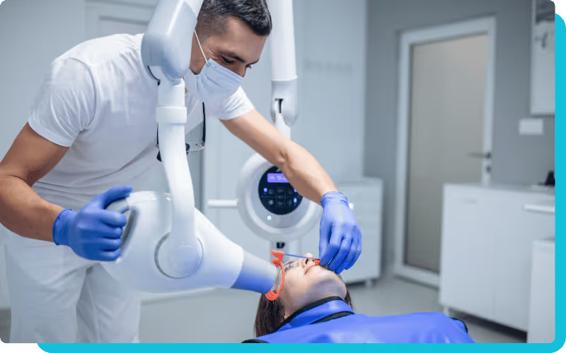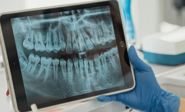
Detecting dental caries early is a fundamental diagnostic challenge. When you identify demineralization and decay in their earliest forms, you directly impact treatment outcomes, patient comfort, and long-term oral health. Yet traditional visual and radiographic interpretation methods often face limitations: sensitivity is lower for subtle lesions, consistency can vary among clinicians, and recognizing early-stage disease can be challenging.
That’s where Pearl AI transforms caries detection. By applying sophisticated algorithms to radiographic images, the system identifies subtle radiographic changes and supports your clinical decision-making with objective, consistent diagnostic intelligence. With Pearl’s pattern-recognition capabilities, you gain enhanced insight into carious activity across surfaces and stages of disease.
Understanding Pearl AI technology for caries detection
Pearl AI uses deep learning and computer vision to review dental radiographs for patterns associated with caries. In the U.S., Second Opinion is FDA-cleared as a computer-aided detection tool for caries on bitewing and periapical radiographs of permanent teeth, used as a second reader. Pearl also received a separate pediatric clearance in 2025 that extends caries support to patients as young as 4 with primary or mixed dentition.
What the system does well is consistency. It processes pixel-level radiographic information, highlights suspected findings with overlays, and helps surface subtle changes that may be easier to miss during routine review. That gives you a more structured radiographic review process without removing clinical judgment from the equation.
What Pearl AI analyzes
- Pixel-level density patterns that may be associated with demineralization or decay
- Anatomical context around contact areas and restorative margins
- Suspected carious findings on supported radiographs, presented with visual overlays
- Radiographic patterns that deserve closer clinical review chairside
When you use the system chairside, you are not just looking at the radiograph alone. You are reviewing it with AI-supported overlays that can help you catch subtle changes earlier, confirm what you already see, and communicate findings more clearly to patients.
Stages of caries development Pearl AI identifies
Pearl AI supports caries detection across a range of radiographic presentations, from subtle early changes to more obvious lesions and recurrent decay around restorations. The value is not that it replaces your diagnosis, but that it helps you review each stage more consistently and with more confidence.
Initial enamel demineralization and white-spot lesions
The earliest radiographic signs of caries can be easy to miss. Slight radiolucency or minimal density change in enamel may not stand out during a fast review. Pearl AI helps flag these subtle patterns so you can take a closer look and consider earlier preventive intervention when appropriate. Clinical correlation still matters, but catching these changes sooner can influence treatment timing.
Enamel caries progression and dentinal involvement
As lesions progress toward or through the dentino-enamel junction, treatment decisions often shift.
Pearl AI can help surface suspicious radiographic changes that suggest a lesion may no longer be confined to enamel, providing stronger support when deciding whether to monitor, remineralize, or restore.
Dentinal caries and pulpal proximity assessment
With deeper lesions, the stakes are higher, but radiographic AI still needs to be interpreted in context. Pearl AI can help identify suspicious dentinal radiolucencies, but final assessment of lesion severity, pulpal risk, and treatment approach still depends on your full clinical evaluation. That is especially important because AI support tools are designed to aid review, not determine severity on their own.
Proximal surface caries in contact areas
Interproximal lesions remain some of the easiest to miss because of overlap and limited direct visibility. This is one of the areas where AI assistance can be especially helpful. Pearl AI adds a second layer of review in contact zones, helping you identify radiographic patterns that may deserve closer attention before the lesion becomes more extensive.
Occlusal caries in pits and fissures
Occlusal surfaces can be difficult to interpret radiographically because of anatomy and superimposition. AI will not solve every occlusal diagnostic challenge, but it can help identify subtle density changes that support a more careful review of suspicious areas. Used alongside your exam and risk assessment, this can strengthen early detection.
Root surface caries and cervical lesions
Root and cervical lesions can be easily underdiagnosed, especially in patients with recession or exposed root surfaces. Pearl AI can help flag suspicious radiographic changes in these zones when they are visible on the image, providing a stronger clinical prompt for evaluation. This can be particularly useful in older adult populations where root caries risk is higher.
Secondary caries around existing restorations
Recurrent caries around restorations remains a common diagnostic challenge because margins, restorative materials, and anatomy can complicate interpretation.
Pearl AI can support review of suspicious radiographic changes adjacent to restoration margins, helping you document and discuss areas that warrant closer clinical examination. Detection still depends on image quality, radiographic visibility, and lesion size.
Clinical applications and diagnostic workflow integration
Here’s how you can use Pearl AI in your everyday workflow to support caries detection, treatment planning, and quality assurance.
Initial patient examination
During your new-patient or comprehensive exam, Pearl AI runs in the background and provides you with annotated radiographs (bitewings, periapicals) that highlight potential carious lesions across every tooth surface. This gives you a baseline diagnostic screen, helps establish thorough documentation, and supports your conversation with the patient on areas of concern.
Recall examination and disease monitoring
For recall visits, you can leverage the AI to compare current and prior radiographs, track lesion progression (or stability), and detect any new caries activity. This gives you objective evidence to monitor changes over time, validate your preventive treatments, and identify new lesions early.
Treatment planning and case presentation
When explaining treatment options to a patient, you’ll appreciate how Pearl AI’s visuals and quantitative data support your recommendations. You can use the flagged areas and overlay annotations to illustrate the carious process, show how early intervention can save tooth structure, and document your findings for internal records and insurance communication.
Quality assurance
AI-assisted detection provides a consistent diagnostic standard across clinicians, regardless of experience level or different chairs or sites. You can use Pearl AI to reduce variability, ensure that no zone is overlooked, and support your practice’s quality-assurance programmes. This helps you maintain diagnostic rigor and standardize your caries-detection protocols.
Advantages of AI-assisted caries detection over traditional methods
Here’s how using Pearl AI gives you distinct advantages compared with standard visual and radiographic interpretation alone.
Enhanced detection of early-stage lesions
With Pearl AI, you gain improved sensitivity for signs of incipient carious lesions such as subtle enamel demineralization and early dentinal involvement. The algorithm picks up density gradients, pixel-level cues, and anatomical context that are harder to detect with the naked eye. Modern AI systems can achieve an accuracy rate of over 90% in detecting dental caries.
This earlier detection allows you to intervene sooner, preserve more tooth structure, and potentially shift treatment toward prevention rather than restoration.
Reduced false negatives and missed diagnoses
A key challenge in caries diagnostics is the risk of overlooking lesions, especially in contact areas, root surfaces, or beneath restorations. With Pearl AI acting as a “second pair of eyes”, you reduce interpretation variability and human perceptual error. Pearl’s deep-learning approach picks up hard-to-spot issues like incipient caries or the early signs of a periapical radiolucency.
This gives you more confidence that diagnostic gaps are minimized.
Objective measurement and documentation
Pearl AI doesn’t just flag areas of concern. With Practice Intelligence, you can surface patients with unscheduled care needs and use AI-supported visuals to aid documentation and communication.
That means you have standardized reporting, improved documentation of findings, and better material for treatment discussions with patients or communications with insurers.
Consistent interpretation across practitioners
In a multi-clinician practice or a group-practice environment, interpretation standards vary. Pearl AI ensures consistent algorithmic assessment: the system will deliver the same high-quality analysis every time.
That consistency helps you maintain diagnostic quality, regardless of operator experience level or fatigue.
Clinical evidence and validation studies
Before implementing any diagnostic aid, you want to understand how well it’s been validated. Here’s what the literature says about Pearl AI.
In a large multi-site study of 8,700+ bitewing and periapical radiographs, Pearl’s AI system showed higher accuracy and consistency for caries detection than unaided dentists.
In general, peer-reviewed research supports that AI software enhances sensitivity and specificity in caries detection. For example, one study showed that Second Opinion improved diagnostic accuracy for caries from 82% to 98% when used by operators.
What this means for you: when you adopt Pearl AI, you’re leveraging a tool grounded in evidence showing that algorithm-based detection can outperform unaided visual or radiographic examination, especially in early-stage and subtle lesions.
Pearl AI implementation guidelines for clinical practices
Here are the practical areas to focus on when integrating Pearl AI into everyday radiographic review.
Technical setup
Start by confirming that your imaging environment is compatible with Pearl. In the U.S., current FDA-cleared caries support applies to bitewing and periapical radiographs, with pediatric support now available for younger patients as well. Before launch, confirm integration requirements, data security expectations, and how results will appear inside your existing imaging workflow.
Staff training and adoption
Training should focus on interpretation, not just software clicks. Your team needs to understand what the overlays represent, how to review flagged areas with clinical context, and how to explain AI-supported findings to patients. The most effective use of Pearl happens when clinicians treat it as a second reader that supports judgment, not replaces it.
Workflow integration
For most practices, the simplest workflow is to incorporate AI into the standard radiographic review process. Capture the image, review the AI-supported overlay, compare with prior images when relevant, then finalize your diagnosis and patient communication. Used this way, Pearl becomes part of a more consistent radiographic review routine rather than a separate extra step.
Quality assurance and continuous review
Pearl AI can also support internal consistency. Reviewing flagged findings across clinicians and over time can help standardize how the practice approaches radiographic interpretation. False positives and false negatives still need to be understood clinically, but using AI findings as part of quality review can strengthen calibration across the team.
Final Thoughts
Dental caries detection has always depended on a combination of clinical skill, radiographic interpretation, and experience. Pearl AI strengthens that process by adding a more consistent, AI-supported review layer that can help surface subtle findings, standardize interpretation, and support clearer patient communication.
Its value is especially clear in the areas where routine review is most variable, such as early lesions, interproximal findings, and suspicious changes around restorations. For practices focused on earlier intervention, stronger documentation, and more consistent radiographic review, Pearl AI offers a practical tool that fits into existing workflows without replacing the clinician’s role.
FAQs
How accurate is Pearl AI compared to traditional caries detection methods?
Pearl’s FDA-cleared AI supports caries detection on 2D bitewing and periapical images and has shown higher accuracy and consistency than unaided dentists in a large multi-site study. Clinical image quality and context still govern outcomes.
Can Pearl AI detect caries on all tooth surfaces?
Pearl AI detects caries across enamel and dentin and on proximal, occlusal, and root surfaces, as well as around restoration margins. Its deep learning algorithms are trained to identify carious activity across the full range of lesion types and locations.
Can Pearl AI differentiate between active and arrested caries?
Not on its own. Pearl AI identifies radiographic patterns that may be consistent with carious lesions, but determining whether a lesion is active or arrested still requires clinical correlation, including visual assessment, patient history, caries risk, and other findings. AI should be used as one part of a full diagnostic evaluation.
Does AI caries detection work with all types of dental X-rays?
In the US, Pearl’s FDA-cleared 2D AI supports bitewing and periapical radiographs for permanent teeth, with a pediatric clearance enabling caries detection in children as young as 4. Panoramic images are supported outside the US. Second Opinion 3D is FDA-cleared to identify anatomy on CBCT, not caries.
What training is needed to use Pearl AI for caries detection?
Using Pearl AI requires only basic orientation on its interface and review protocols. Once it is integrated into your workflow, you can interpret its overlays alongside your radiographs, review flagged findings, and use them to support diagnostic accuracy and quality assurance.