Artificial intelligence is raising the bar for dental radiographic analysis. By reading pixel patterns, density shifts, and anatomical relationships at scale, AI systems can surface early and subtle findings that traditional visual interpretation often overlooks. Recent systematic reviews and clinical studies report strong performance for AI in detecting caries and periapical radiolucencies across intraoral, panoramic, and periapical images.
In practice, AI does not replace your judgment. It adds a consistent, quantitative layer that enhances accuracy, speeds up the review process, and supports clearer treatment discussions. When you combine AI’s pattern recognition with your clinical context, you can catch problems earlier and plan with greater precision.
Understanding AI capabilities in dental radiographic analysis
AI systems analyze dental X-rays using algorithms that evaluate pixel intensities, edges, textures, and geometric relationships with mathematical precision. Trained on thousands of annotated images, these models learn to recognize patterns linked to disease, from incipient enamel changes to apical radiolucencies.
Because the models are consistent and tireless, they reduce variability that can arise from human factors. Studies show AI can match or exceed specialist-level accuracy in tasks like caries detection and periapical assessment, particularly when applied to standardized radiographic inputs.
What human eyes miss in dental X-ray interpretation
Even experienced clinicians face natural constraints when reading large volumes of radiographs. AI helps counter these limitations by staying consistent across cases and time.
Visual perception limits and fatigue
Sustained reading can reduce attention and sensitivity to low-contrast details. AI maintains stable performance across long sessions and complex cases, which supports more consistent detection.
Subtle density changes and early pathology
Minor density variations that signal early caries or early periapical change can fall below human detection thresholds. AI models trained on pixel-level patterns improve sensitivity to these incipient signals.
Overlaps, superimposition, and hard-to-see regions
Superimposed anatomy and complex spatial relationships can obscure pathology on 2D images. Object-detection and segmentation models help separate structures and highlight suspicious regions, improving visibility in challenging areas.
Inconsistent interpretation and cognitive load
Experience, training background, and case pressure can lead to variation across readers. By applying learned criteria uniformly, AI reduces inter-reader variability and supports standardized assessments.
Time pressure and multi-case review
High-throughput environments increase the risk of oversight. Automated prescreening and prioritization can flag likely positives for closer review, helping you focus time where it matters most.
Rare pattern recognition and measurement precision
Uncommon presentations and small dimensional differences can be easily missed. AI brings quantitative measurements and consistent scoring, which supports recognition of atypical patterns and more precise comparisons over time.
How AI finds what eyes miss: core detection methods
AI-powered radiographic systems use advanced algorithms to uncover subtle details and patterns that traditional visual inspection often overlooks. By combining deep learning, computer vision, and mathematical modeling, these tools can evaluate every pixel in a radiograph with precision that exceeds the human eye.
Multi-dimensional pattern recognition
Unlike the human eye, which primarily interprets 2D patterns, AI can process data in multiple dimensions, accounting for grayscale gradients, spatial geometry, and density variations. Deep-learning segmentation models have demonstrated a strong ability to map bone and soft tissue boundaries in panoramic and CBCT images, thereby improving diagnostic consistency across complex cases.
Quantitative measurement and standardized scoring
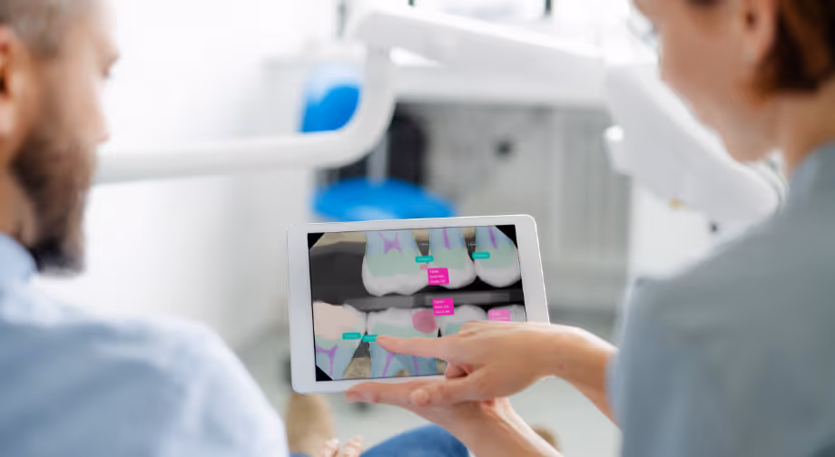
AI provides exact measurements and objective scoring based on mathematical criteria, removing subjective variability. Systems like Pearl’s Second Opinion deliver standardized lesion detection and quantification, helping you track clinical changes more precisely and maintain consistent quality assurance across providers.
Longitudinal and comparative change detection
Machine learning can compare current and past radiographs, highlighting subtle differences over time that indicate disease progression or treatment response.
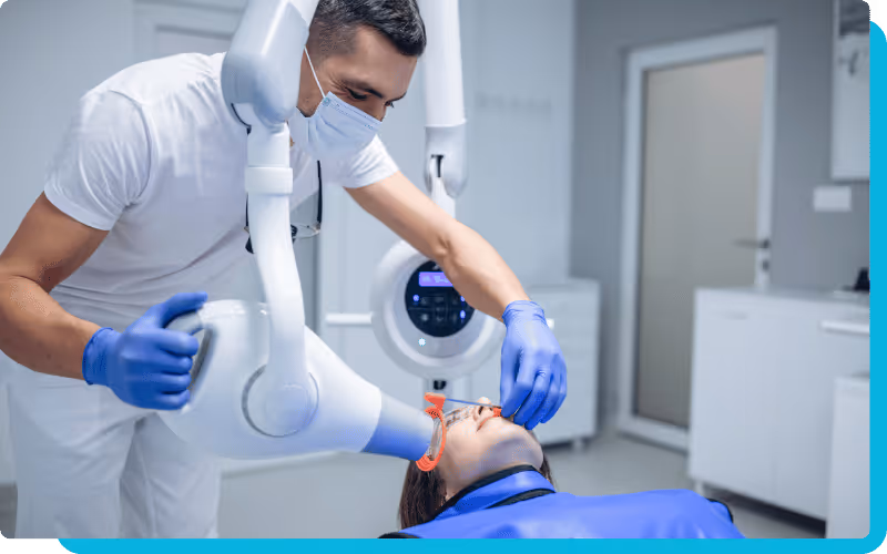
Real-time image quality checks and retake guidance
AI systems can instantly assess exposure, contrast, and positioning, flagging low-quality images before they are interpreted. This helps you avoid diagnostic uncertainty by prompting timely retakes.
Periodontal and bone level assessment
Algorithms can quantify bone levels and measure attachment loss in a fraction of the time it takes to do so manually. These measurements help you monitor progression and tailor treatment plans more effectively.
Caries, periapical, and root morphology flagging
AI highlights early carious lesions, periapical pathology, and root morphology variations that may be subtle or obscured by overlapping anatomy. This ensures that potential issues are brought to your attention early, reducing missed diagnoses and supporting preventive care.
Clinical benefits of AI-enhanced radiographic analysis
Integrating AI into your radiographic workflow delivers measurable clinical benefits. It supports earlier disease detection, increases diagnostic reliability, and enhances communication with both patients and colleagues.
Earlier disease detection and intervention
AI enables you to identify caries, endodontic pathology, or bone loss at an earlier stage than would typically be possible through visual examination alone. This supports preventive treatment approaches and better long-term outcomes.
Fewer misses and more consistent calls
AI minimizes the impact of fatigue, bias, and time pressure on diagnostic accuracy. It acts as a second reviewer, ensuring that key findings are not overlooked. This results in fewer missed diagnoses and higher confidence in clinical decision-making.
Standardized interpretation across providers
Machine learning applies the same detection criteria to every case, creating consistent interpretation standards across different clinicians. This is particularly useful in multi-dentist or multi-location practices, where diagnostic variability can be a challenge.
Better treatment planning and risk communication
AI-generated reports provide objective data that support risk discussions and treatment explanations with patients. Visual overlays on radiographs make it easier for patients to understand the findings, which increases trust and treatment acceptance.
Implementation best practices for AI radiographic systems
Introducing AI dental X-ray analysis into your practice is most effective when approached strategically. You should begin by selecting validated tools, training your team, and integrating systems into existing workflows.
Steps for successful implementation:
- Select clinically validated AI tools: Choose systems that have regulatory clearance (FDA, CE, or local equivalent) and peer-reviewed evidence of diagnostic accuracy.
- Start with a pilot deployment: Implement the system in a limited scope to measure accuracy, clinician satisfaction, and workflow impact before rolling it out fully.
- Train your staff thoroughly: Educate clinicians and assistants on how to interpret AI results, verify findings, and document their use appropriately.
- Maintain quality assurance: Periodically audit AI results against human assessments to ensure consistent performance and reliability.
- Integrate with existing imaging systems: Use software that connects seamlessly with your radiography, EHR, and practice management systems to minimize workflow disruption.
When used correctly, AI becomes a clinical partner that enhances rather than replaces human expertise, helping you improve diagnostic quality and patient outcomes.
How Pearl’s AI enhances radiographic accuracy
Pearl’s FDA-approved, AI-powered solutions help you achieve greater precision in every stage of radiographic analysis. Second Opinion delivers real-time pathology detection, flagging subtle findings that may be difficult to see with the human eye. Paired with ImageCheck, Pearl’s new real-time X-ray quality assurance platform, you can ensure every image meets diagnostic standards before review, reducing retakes and improving consistency across your workflow.
Together, these tools enhance diagnostic accuracy, streamline imaging processes, and strengthen patient confidence in your care.
FAQs
What types of dental conditions can AI detect that humans might miss?
AI can identify early-stage caries, periapical pathology, bone density loss, impacted teeth, and root anomalies, conditions that are sometimes below the human threshold for visual detection.
Can AI completely replace human radiographic interpretation?
No. AI is designed to support, not replace, your expertise. It offers a second, objective review that improves accuracy and consistency while leaving the final diagnosis to you.
What training is needed for dental staff to use AI X-ray analysis?
Training should include interpreting AI-generated findings, verifying flagged results, and understanding how AI integrates with imaging workflows. Vendors often provide onboarding sessions and clinical resources to support their clients.
How does AI improve early detection of dental diseases?
AI evaluates pixel-level patterns that indicate disease long before visible symptoms appear, enabling preventive intervention and reducing long-term complications.